Serial Femtosecond Crystallography and Ultrafast Absorption Spectroscopy of the Photoswitchable Fluorescent Protein Irisfp.
Colletier, J., Sliwa, M., Gallat, F., Sugahara, M., Guillon, V., Schiro, G., Coquelle, N., Woodhouse, J., Roux, L., Gotthard, G., Royant, A., Uriarte, L.M., Ruckebusch, C., Joti, Y., Byrdin, M., Mizohata, E., Nango, E., Tanaka, T., Tono, K., Yabashi, M., Adam, V., Cammarata, M., Schlichting, I., Bourgeois, D., Weik, M.(2016) J Phys Chem 7: 882
- PubMed: 26866390 Search on PubMed
- DOI: https://doi.org/10.1021/acs.jpclett.5b02789
- Primary Citation Related Structures:
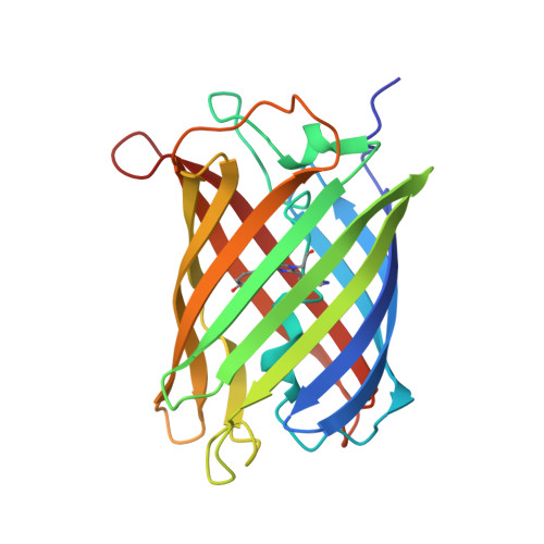
5FVF, 5FVG, 5FVI - PubMed Abstract:
Reversibly photoswitchable fluorescent proteins find growing applications in cell biology, yet mechanistic details, in particular on the ultrafast photochemical time scale, remain unknown. We employed time-resolved pump-probe absorption spectroscopy on the reversibly photoswitchable fluorescent protein IrisFP in solution to study photoswitching from the nonfluorescent (off) to the fluorescent (on) state. Evidence is provided for the existence of several intermediate states on the pico- and microsecond time scales that are attributed to chromophore isomerization and proton transfer, respectively. Kinetic modeling favors a sequential mechanism with the existence of two excited state intermediates with lifetimes of 2 and 15 ps, the second of which controls the photoswitching quantum yield. In order to support that IrisFP is suited for time-resolved experiments aiming at a structural characterization of these ps intermediates, we used serial femtosecond crystallography at an X-ray free electron laser and solved the structure of IrisFP in its on state. Sample consumption was minimized by embedding crystals in mineral grease, in which they remain photoswitchable. Our spectroscopic and structural results pave the way for time-resolved serial femtosecond crystallography aiming at characterizing the structure of ultrafast intermediates in reversibly photoswitchable fluorescent proteins.
- Institut de Biologie Structurale , Université de Grenoble Alpes, CEA, CNRS, F-38044 Grenoble, France.
Organizational Affiliation: