5FM5 | pdb_00005fm5

Crystal structure of the myomesin:obscurin-like-1 complex


Experimental Data Snapshot

  • Method: X-RAY DIFFRACTION
  • Resolution: 3.10 Å
  • R-Value Free: 
    0.259 (Depositor), 0.257 (DCC) 
  • R-Value Work: 
    0.216 (Depositor), 0.216 (DCC) 
  • R-Value Observed: 
    0.218 (Depositor) 

Starting Models: experimental
View more details

wwPDB Validation 3D Report Full Report

Validation slider image for 5FM5

This is version 1.4 of the entry. See complete history

Literature

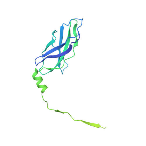
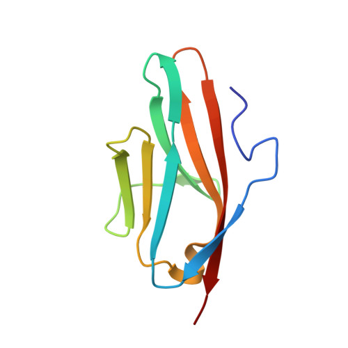
Binding of Myomesin to Obscurin-Like-1 at the Muscle M-Band Provides a Strategy for Isoform-Specific Mechanical Protection.

Pernigo, S.Fukuzawa, A.Beedle, A.E.Holt, M.Round, A.Pandini, A.Garcia-Manyes, S.Gautel, M.Steiner, R.A.

(2017) Structure 25: 107-120

  • DOI: https://doi.org/10.1016/j.str.2016.11.015
  • Primary Citation Related Structures: 
    5FM4, 5FM5, 5FM8

  • PubMed Abstract: 

    The sarcomeric cytoskeleton is a network of modular proteins that integrate mechanical and signaling roles. Obscurin, or its homolog obscurin-like-1, bridges the giant ruler titin and the myosin crosslinker myomesin at the M-band. Yet, the molecular mechanisms underlying the physical obscurin(-like-1):myomesin connection, important for mechanical integrity of the M-band, remained elusive. Here, using a combination of structural, cellular, and single-molecule force spectroscopy techniques, we decode the architectural and functional determinants defining the obscurin(-like-1):myomesin complex. The crystal structure reveals a trans-complementation mechanism whereby an incomplete immunoglobulin-like domain assimilates an isoform-specific myomesin interdomain sequence. Crucially, this unconventional architecture provides mechanical stability up to forces of ∼135 pN. A cellular competition assay in neonatal rat cardiomyocytes validates the complex and provides the rationale for the isoform specificity of the interaction. Altogether, our results reveal a novel binding strategy in sarcomere assembly, which might have implications on muscle nanomechanics and overall M-band organization.


  • Organizational Affiliation
    • Randall Division of Cell and Molecular Biophysics, King's College London, London SE1 1UL, UK.

Macromolecule Content 

  • Total Structure Weight: 73.02 kDa 
  • Atom Count: 3,566 
  • Modeled Residue Count: 441 
  • Deposited Residue Count: 658 
  • Unique protein chains: 2

Macromolecules

Find similar proteins by:|  3D Structure
Entity ID: 1
MoleculeChains  Sequence LengthOrganismDetailsImage
MYOMESIN-1A [auth M],
B [auth N]
231Homo sapiensMutation(s): 0 
UniProt & NIH Common Fund Data Resources
Find proteins for P52179 (Homo sapiens)
Explore P52179 
Go to UniProtKB:  P52179
PHAROS:  P52179
GTEx:  ENSG00000101605 
Entity Groups
Sequence Clusters30% Identity50% Identity70% Identity90% Identity95% Identity100% Identity
UniProt GroupP52179
Sequence Annotations
Expand
Reference Sequence
Find similar proteins by:|  3D Structure
Entity ID: 2
MoleculeChains  Sequence LengthOrganismDetailsImage
OBSCURIN-LIKE-1C [auth O],
D [auth P]
98Homo sapiensMutation(s): 0 
UniProt & NIH Common Fund Data Resources
Find proteins for O75147 (Homo sapiens)
Explore O75147 
Go to UniProtKB:  O75147
PHAROS:  O75147
GTEx:  ENSG00000124006 
Entity Groups
Sequence Clusters30% Identity50% Identity70% Identity90% Identity95% Identity100% Identity
UniProt GroupO75147
Sequence Annotations
Expand
Reference Sequence

Experimental Data & Validation

Experimental Data

  • Method: X-RAY DIFFRACTION
  • Resolution: 3.10 Å
  • R-Value Free:  0.259 (Depositor), 0.257 (DCC) 
  • R-Value Work:  0.216 (Depositor), 0.216 (DCC) 
  • R-Value Observed: 0.218 (Depositor) 
Space Group: C 1 2 1
Unit Cell:
Length ( Å )Angle ( ˚ )
a = 91.18α = 90
b = 134.38β = 92.5
c = 68.39γ = 90
Software Package:
Software NamePurpose
XDSdata reduction
Aimlessdata scaling
PHASERphasing
MOLREPphasing
REFMACrefinement

Structure Validation

View Full Validation Report


Entry History 

Deposition Data

Revision History  (Full details and data files)

  • Version 1.0: 2016-11-23
    Type: Initial release
  • Version 1.1: 2016-12-07
    Changes: Database references
  • Version 1.2: 2016-12-28
    Changes: Database references
  • Version 1.3: 2017-03-01
    Changes: Database references
  • Version 1.4: 2024-01-10
    Changes: Data collection, Database references, Other, Refinement description