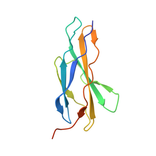
5FM4

Structure of the C-terminally extended domain My4 of human myomesin (space group P21)


Experimental Data Snapshot

  • Method: X-RAY DIFFRACTION
  • Resolution: 2.80 Å
  • R-Value Free: 0.287 
  • R-Value Work: 0.261 
  • R-Value Observed: 0.262 

Starting Model: experimental
View more details

wwPDB Validation   3D Report Full Report


This is version 2.1 of the entry. See complete history


Literature

Binding of Myomesin to Obscurin-Like-1 at the Muscle M-Band Provides a Strategy for Isoform-Specific Mechanical Protection.

Pernigo, S.Fukuzawa, A.Beedle, A.E.Holt, M.Round, A.Pandini, A.Garcia-Manyes, S.Gautel, M.Steiner, R.A.

(2017) Structure 25: 107-120

  • DOI: https://doi.org/10.1016/j.str.2016.11.015
  • Primary Citation of Related Structures:  
    5FM4, 5FM5, 5FM8

  • PubMed Abstract: 

    The sarcomeric cytoskeleton is a network of modular proteins that integrate mechanical and signaling roles. Obscurin, or its homolog obscurin-like-1, bridges the giant ruler titin and the myosin crosslinker myomesin at the M-band. Yet, the molecular mechanisms underlying the physical obscurin(-like-1):myomesin connection, important for mechanical integrity of the M-band, remained elusive. Here, using a combination of structural, cellular, and single-molecule force spectroscopy techniques, we decode the architectural and functional determinants defining the obscurin(-like-1):myomesin complex. The crystal structure reveals a trans-complementation mechanism whereby an incomplete immunoglobulin-like domain assimilates an isoform-specific myomesin interdomain sequence. Crucially, this unconventional architecture provides mechanical stability up to forces of ∼135 pN. A cellular competition assay in neonatal rat cardiomyocytes validates the complex and provides the rationale for the isoform specificity of the interaction. Altogether, our results reveal a novel binding strategy in sarcomere assembly, which might have implications on muscle nanomechanics and overall M-band organization.


  • Organizational Affiliation

    Randall Division of Cell and Molecular Biophysics, King's College London, London SE1 1UL, UK.


Macromolecules
Find similar proteins by:  (by identity cutoff)  |  3D Structure
Entity ID: 1
MoleculeChains Sequence LengthOrganismDetailsImage
MYOMESIN-1
A, B, C, D, E
A, B, C, D, E, F
112Homo sapiensMutation(s): 0 
UniProt & NIH Common Fund Data Resources
Find proteins for P52179 (Homo sapiens)
Explore P52179 
Go to UniProtKB:  P52179
PHAROS:  P52179
GTEx:  ENSG00000101605 
Entity Groups  
Sequence Clusters30% Identity50% Identity70% Identity90% Identity95% Identity100% Identity
UniProt GroupP52179
Sequence Annotations
Expand
  • Reference Sequence
Experimental Data & Validation

Experimental Data

  • Method: X-RAY DIFFRACTION
  • Resolution: 2.80 Å
  • R-Value Free: 0.287 
  • R-Value Work: 0.261 
  • R-Value Observed: 0.262 
  • Space Group: P 1 21 1
Unit Cell:
Length ( Å )Angle ( ˚ )
a = 43.53α = 90
b = 156.84β = 94.38
c = 48.26γ = 90
Software Package:
Software NamePurpose
PHENIXrefinement
XDSdata reduction
Aimlessdata scaling
PHASERphasing

Structure Validation

View Full Validation Report


Entry History 

Deposition Data

Revision History  (Full details and data files)

  • Version 1.0: 2016-11-23
    Type: Initial release
  • Version 1.1: 2016-12-07
    Changes: Database references
  • Version 1.2: 2016-12-28
    Changes: Database references
  • Version 1.3: 2017-03-01
    Changes: Database references
  • Version 2.0: 2019-10-23
    Changes: Atomic model, Data collection, Other
  • Version 2.1: 2024-01-10
    Changes: Data collection, Database references, Refinement description