Structural Insights into the Distinct Binding Mode of Cyclic Di-AMP with SaCpaA_RCK.
Chin, K.H., Liang, J.M., Yang, J.G., Shih, M.S., Tu, Z.L., Wang, Y.C., Sun, X.H., Hu, N.J., Liang, Z.X., Dow, J.M., Ryan, R.P., Chou, S.H.(2015) Biochemistry 54: 4936-4951
- PubMed: 26171638 Search on PubMed
- DOI: https://doi.org/10.1021/acs.biochem.5b00633
- Primary Citation Related Structures:
4YP1, 5F29 - PubMed Abstract:
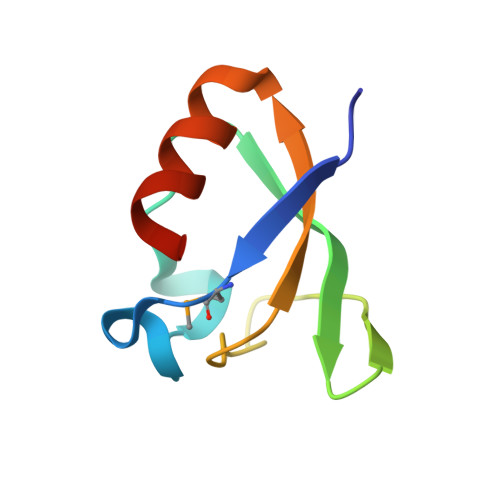
Cyclic di-AMP (c-di-AMP) is a relatively new member of the family of bacterial cyclic dinucleotide second messengers. It has attracted significant attention in recent years because of the abundant roles it plays in a variety of Gram-positive bacteria. The structural features that allow diverse bacterial proteins to bind c-di-AMP are not fully understood. Here we report the biophysical and structural studies of c-di-AMP in complex with a bacterial cation-proton antiporter (CpaA) RCK (regulator of the conductance of K(+)) protein from Staphylococcus aureus (Sa). The crystal structure of the SaCpaA_RCK C-terminal domain (CTD) in complex with c-di-AMP was determined to a resolution of 1.81 Å. This structure revealed two well-liganded water molecules, each interacting with one of the adenine bases by a unique H2Olp-π interaction to stabilize the complex. Sequence blasting using the SaCpaA_RCK primary sequence against the bacterial genome database returned many CpaA analogues, and alignment of these sequences revealed that the active site residues are all well-conserved, indicating a universal c-di-AMP binding mode for CpaA_RCK. A proteoliposome activity assay using the full-length SaCpaA membrane protein indicated that c-di-AMP binding alters its antiporter activity by approximately 40%. A comparison of this structure to all other reported c-di-AMP-receptor complex structures revealed that c-di-AMP binds to receptors in either a "U-shape" or "V-shape" mode. The two adenine rings are stabilized in the inner interaction zone by a variety of CH-π, cation-π, backbone-π, or H2Olp-π interaction, but more commonly in the outer interaction zone by hydrophobic CH-π or π-π interaction. The structures determined to date provide an understanding of the mechanisms by which a single c-di-AMP can interact with a variety of receptor proteins, and how c-di-AMP binds receptor proteins in a special way different from that of c-di-GMP.
- †National Chung Hsing University Biotechnology Center, National Chung Hsing University, Taichung 40227, Taiwan, ROC.
Organizational Affiliation: