5F0M

Structure of retromer VPS26-VPS35 subunits bound to SNX3 and DMT1 (SeMet labeled)


Experimental Data Snapshot

  • Method: X-RAY DIFFRACTION
  • Resolution: 3.10 Å
  • R-Value Free: 0.254 
  • R-Value Work: 0.210 
  • R-Value Observed: 0.212 

wwPDB Validation   3D Report Full Report


This is version 1.2 of the entry. See complete history


Literature

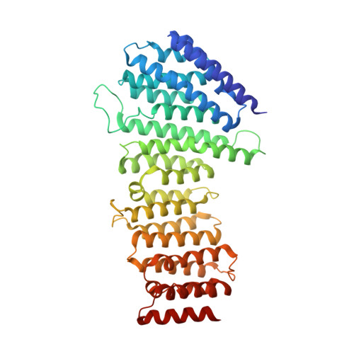
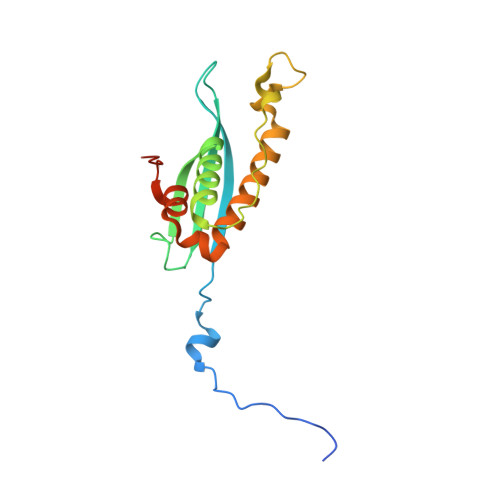
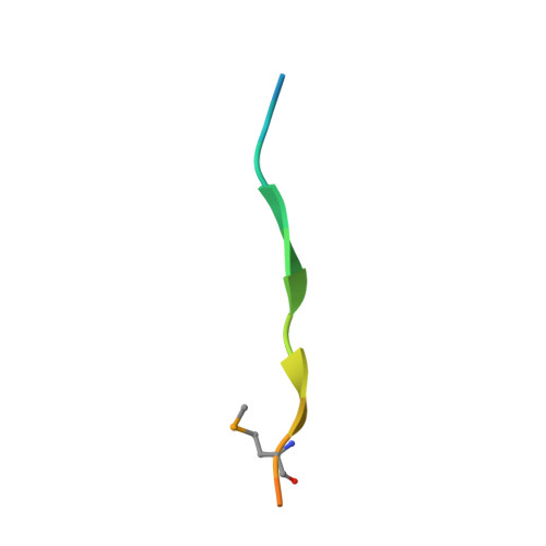
Structural Mechanism for Cargo Recognition by the Retromer Complex.

Lucas, M.Gershlick, D.C.Vidaurrazaga, A.Rojas, A.L.Bonifacino, J.S.Hierro, A.

(2016) Cell 167: 1623-1635.e14

  • DOI: https://doi.org/10.1016/j.cell.2016.10.056
  • Primary Citation of Related Structures:  
    5F0J, 5F0K, 5F0L, 5F0M, 5F0P

  • PubMed Abstract: 

    Retromer is a multi-protein complex that recycles transmembrane cargo from endosomes to the trans-Golgi network and the plasma membrane. Defects in retromer impair various cellular processes and underlie some forms of Alzheimer's disease and Parkinson's disease. Although retromer was discovered over 15 years ago, the mechanisms for cargo recognition and recruitment to endosomes have remained elusive. Here, we present an X-ray crystallographic analysis of a four-component complex comprising the VPS26 and VPS35 subunits of retromer, the sorting nexin SNX3, and a recycling signal from the divalent cation transporter DMT1-II. This analysis identifies a binding site for canonical recycling signals at the interface between VPS26 and SNX3. In addition, the structure highlights a network of cooperative interactions among the VPS subunits, SNX3, and cargo that couple signal-recognition to membrane recruitment.


  • Organizational Affiliation

    Structural Biology Unit, CIC bioGUNE, Bizkaia Technology Park, 48160 Derio, Spain.


Macromolecules
Find similar proteins by:  (by identity cutoff)  |  3D Structure
Entity ID: 1
MoleculeChains Sequence LengthOrganismDetailsImage
Vacuolar protein sorting-associated protein 35462Homo sapiensMutation(s): 0 
Gene Names: VPS35MEM3TCCCTA00141
UniProt & NIH Common Fund Data Resources
Find proteins for Q96QK1 (Homo sapiens)
Explore Q96QK1 
Go to UniProtKB:  Q96QK1
PHAROS:  Q96QK1
GTEx:  ENSG00000069329 
Entity Groups  
Sequence Clusters30% Identity50% Identity70% Identity90% Identity95% Identity100% Identity
UniProt GroupQ96QK1
Sequence Annotations
Expand
  • Reference Sequence
Find similar proteins by:  (by identity cutoff)  |  3D Structure
Entity ID: 2
MoleculeChains Sequence LengthOrganismDetailsImage
Vacuolar protein sorting-associated protein 26A321Homo sapiensMutation(s): 0 
Gene Names: VPS26AVPS26
UniProt & NIH Common Fund Data Resources
Find proteins for O75436 (Homo sapiens)
Explore O75436 
Go to UniProtKB:  O75436
PHAROS:  O75436
GTEx:  ENSG00000122958 
Entity Groups  
Sequence Clusters30% Identity50% Identity70% Identity90% Identity95% Identity100% Identity
UniProt GroupO75436
Sequence Annotations
Expand
  • Reference Sequence
Find similar proteins by:  (by identity cutoff)  |  3D Structure
Entity ID: 3
MoleculeChains Sequence LengthOrganismDetailsImage
Sorting nexin-3167Homo sapiensMutation(s): 0 
Gene Names: SNX3
UniProt & NIH Common Fund Data Resources
Find proteins for O60493 (Homo sapiens)
Explore O60493 
Go to UniProtKB:  O60493
PHAROS:  O60493
GTEx:  ENSG00000112335 
Entity Groups  
Sequence Clusters30% Identity50% Identity70% Identity90% Identity95% Identity100% Identity
UniProt GroupO60493
Sequence Annotations
Expand
  • Reference Sequence

Find similar proteins by:  Sequence   |   3D Structure  

Entity ID: 4
MoleculeChains Sequence LengthOrganismDetailsImage
Natural resistance-associated macrophage protein 218Homo sapiensMutation(s): 0 
Gene Names: SLC11A2DCT1DMT1NRAMP2OK/SW-cl.20
UniProt & NIH Common Fund Data Resources
Find proteins for P49281 (Homo sapiens)
Explore P49281 
Go to UniProtKB:  P49281
PHAROS:  P49281
GTEx:  ENSG00000110911 
Entity Groups  
Sequence Clusters30% Identity50% Identity70% Identity90% Identity95% Identity100% Identity
UniProt GroupP49281
Sequence Annotations
Expand
  • Reference Sequence
Small Molecules
Ligands 3 Unique
IDChains Name / Formula / InChI Key2D Diagram3D Interactions
SO4
Query on SO4

Download Ideal Coordinates CCD File 
E [auth A]
F [auth A]
M [auth B]
V [auth C]
W [auth C]
E [auth A],
F [auth A],
M [auth B],
V [auth C],
W [auth C],
X [auth C]
SULFATE ION
O4 S
QAOWNCQODCNURD-UHFFFAOYSA-L
GOL
Query on GOL

Download Ideal Coordinates CCD File 
G [auth A],
H [auth A],
N [auth B],
Y [auth C],
Z [auth C]
GLYCEROL
C3 H8 O3
PEDCQBHIVMGVHV-UHFFFAOYSA-N
EDO
Query on EDO

Download Ideal Coordinates CCD File 
AA [auth C]
I [auth A]
J [auth A]
K [auth A]
L [auth A]
AA [auth C],
I [auth A],
J [auth A],
K [auth A],
L [auth A],
O [auth B],
P [auth B],
Q [auth B],
R [auth B],
S [auth B],
T [auth B],
U [auth B]
1,2-ETHANEDIOL
C2 H6 O2
LYCAIKOWRPUZTN-UHFFFAOYSA-N
Modified Residues  1 Unique
IDChains TypeFormula2D DiagramParent
MSE
Query on MSE
B
L-PEPTIDE LINKINGC5 H11 N O2 SeMET
Experimental Data & Validation

Experimental Data

  • Method: X-RAY DIFFRACTION
  • Resolution: 3.10 Å
  • R-Value Free: 0.254 
  • R-Value Work: 0.210 
  • R-Value Observed: 0.212 
  • Space Group: C 1 2 1
Unit Cell:
Length ( Å )Angle ( ˚ )
a = 369.48α = 90
b = 74.91β = 97.64
c = 57.39γ = 90
Software Package:
Software NamePurpose
XSCALEdata scaling
REFMACrefinement
PDB_EXTRACTdata extraction
XDSdata reduction
PHASERphasing

Structure Validation

View Full Validation Report


Entry History & Funding Information

Deposition Data


Funding OrganizationLocationGrant Number
the Carlos III Health InstituteSpainPI11/00121
Basque GovernmentSpainPI2011-26
Spanish Ministry of Economy and CompetitivenessSpainBFU2014-59759-R

Revision History  (Full details and data files)

  • Version 1.0: 2016-12-07
    Type: Initial release
  • Version 1.1: 2016-12-14
    Changes: Database references
  • Version 1.2: 2024-01-10
    Changes: Data collection, Database references, Refinement description