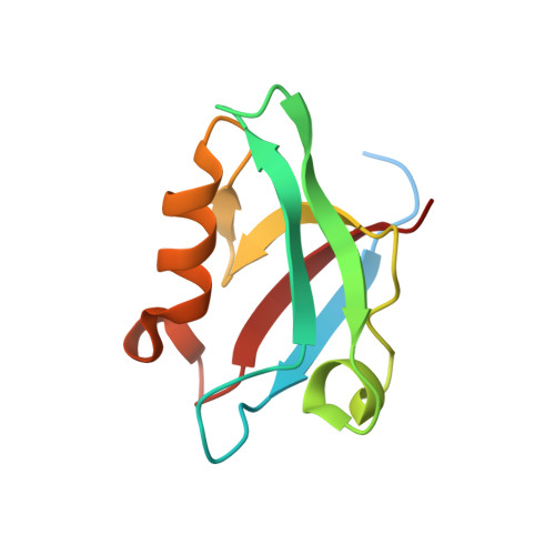
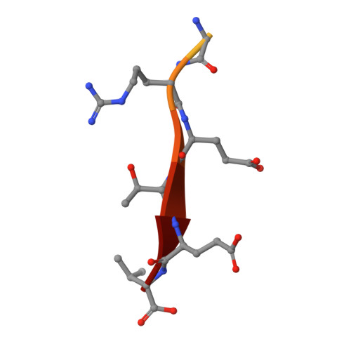
5EYZ

CRYSTAL STRUCTURE OF THE PTPN4 PDZ DOMAIN COMPLEXED WITH THE TAILORED PEPTIDE CYTO8-RETEV


Experimental Data Snapshot

  • Method: X-RAY DIFFRACTION
  • Resolution: 2.09 Å
  • R-Value Free: 0.239 
  • R-Value Work: 0.229 
  • R-Value Observed: 0.230 

Starting Model: experimental
View more details

wwPDB Validation   3D Report Full Report


This is version 1.7 of the entry. See complete history


Literature

Molecular Basis of the Interaction of the Human Protein Tyrosine Phosphatase Non-receptor Type 4 (PTPN4) with the Mitogen-activated Protein Kinase p38 gamma.

Maisonneuve, P.Caillet-Saguy, C.Vaney, M.C.Bibi-Zainab, E.Sawyer, K.Raynal, B.Haouz, A.Delepierre, M.Lafon, M.Cordier, F.Wolff, N.

(2016) J Biol Chem 291: 16699-16708

  • DOI: https://doi.org/10.1074/jbc.M115.707208
  • Primary Citation of Related Structures:  
    5EYZ, 5EZ0

  • PubMed Abstract: 

    The human protein tyrosine phosphatase non-receptor type 4 (PTPN4) prevents cell death induction in neuroblastoma and glioblastoma cell lines in a PDZ·PDZ binding motifs-dependent manner, but the cellular partners of PTPN4 involved in cell protection are unknown. Here, we described the mitogen-activated protein kinase p38γ as a cellular partner of PTPN4. The main contribution to the p38γ·PTPN4 complex formation is the tight interaction between the C terminus of p38γ and the PDZ domain of PTPN4. We solved the crystal structure of the PDZ domain of PTPN4 bound to the p38γ C terminus. We identified the molecular basis of recognition of the C-terminal sequence of p38γ that displays the highest affinity among all endogenous partners of PTPN4. We showed that the p38γ C terminus is also an efficient inducer of cell death after its intracellular delivery. In addition to recruiting the kinase, the binding of the C-terminal sequence of p38γ to PTPN4 abolishes the catalytic autoinhibition of PTPN4 and thus activates the phosphatase, which can efficiently dephosphorylate the activation loop of p38γ. We presume that the p38γ·PTPN4 interaction promotes cellular signaling, preventing cell death induction.


  • Organizational Affiliation

    From the Département de Biologie Structurale et Chimie, Unité de Résonance Magnétique Nucléaire des Biomolécules, UMR 3528 and Université Pierre et Marie Curie, Cellule Pasteur UPMC, 75005 Paris, France.


Macromolecules
Find similar proteins by:  (by identity cutoff)  |  3D Structure
Entity ID: 1
MoleculeChains Sequence LengthOrganismDetailsImage
Tyrosine-protein phosphatase non-receptor type 4
A, B, C, D
107Homo sapiensMutation(s): 0 
Gene Names: PTPN4
EC: 3.1.3.48
UniProt & NIH Common Fund Data Resources
Find proteins for P29074 (Homo sapiens)
Explore P29074 
Go to UniProtKB:  P29074
PHAROS:  P29074
GTEx:  ENSG00000088179 
Entity Groups  
Sequence Clusters30% Identity50% Identity70% Identity90% Identity95% Identity100% Identity
UniProt GroupP29074
Sequence Annotations
Expand
  • Reference Sequence

Find similar proteins by:  Sequence   |   3D Structure  

Entity ID: 2
MoleculeChains Sequence LengthOrganismDetailsImage
CYTO8-RETEV
E, F, G, H
13synthetic constructMutation(s): 0 
Entity Groups  
Sequence Clusters30% Identity50% Identity70% Identity90% Identity95% Identity100% Identity
Sequence Annotations
Expand
  • Reference Sequence
Small Molecules
Ligands 1 Unique
IDChains Name / Formula / InChI Key2D Diagram3D Interactions
CL
Query on CL

Download Ideal Coordinates CCD File 
I [auth C]CHLORIDE ION
Cl
VEXZGXHMUGYJMC-UHFFFAOYSA-M
Experimental Data & Validation

Experimental Data

  • Method: X-RAY DIFFRACTION
  • Resolution: 2.09 Å
  • R-Value Free: 0.239 
  • R-Value Work: 0.229 
  • R-Value Observed: 0.230 
  • Space Group: C 2 2 21
Unit Cell:
Length ( Å )Angle ( ˚ )
a = 77.71α = 90
b = 80.88β = 90
c = 170.23γ = 90
Software Package:
Software NamePurpose
BUSTER-TNTrefinement
SCALAdata scaling
PDB_EXTRACTdata extraction
XDSdata reduction
PHASERphasing

Structure Validation

View Full Validation Report


Entry History 

Deposition Data

Revision History  (Full details and data files)

  • Version 1.0: 2016-06-08
    Type: Initial release
  • Version 1.1: 2016-06-15
    Changes: Database references
  • Version 1.2: 2016-08-17
    Changes: Database references
  • Version 1.3: 2016-10-19
    Changes: Refinement description
  • Version 1.4: 2017-12-20
    Changes: Database references, Structure summary
  • Version 1.5: 2019-10-16
    Changes: Data collection
  • Version 1.6: 2024-01-10
    Changes: Data collection, Database references, Refinement description
  • Version 1.7: 2024-10-16
    Changes: Structure summary