5EJB

Crystal structure of prefusion Hendra virus F protein


Experimental Data Snapshot

  • Method: X-RAY DIFFRACTION
  • Resolution: 3.20 Å
  • R-Value Free: 0.283 
  • R-Value Work: 0.253 
  • R-Value Observed: 0.254 

wwPDB Validation   3D Report Full Report


Ligand Structure Quality Assessment 


This is version 2.1 of the entry. See complete history


Literature

Structure and stabilization of the Hendra virus F glycoprotein in its prefusion form.

Wong, J.J.Paterson, R.G.Lamb, R.A.Jardetzky, T.S.

(2016) Proc Natl Acad Sci U S A 113: 1056-1061

  • DOI: https://doi.org/10.1073/pnas.1523303113
  • Primary Citation of Related Structures:  
    5EJB

  • PubMed Abstract: 

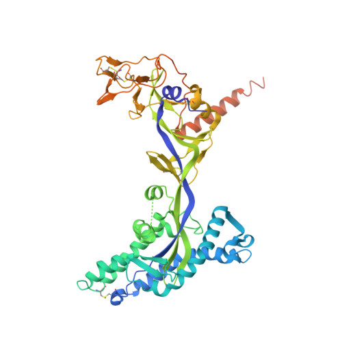
    Hendra virus (HeV) is one of the two prototypical members of the Henipavirus genus of paramyxoviruses, which are designated biosafety level 4 (BSL-4) organisms due to the high mortality rate of Nipah virus (NiV) and HeV in humans. Paramyxovirus cell entry is mediated by the fusion protein, F, in response to binding of a host receptor by the attachment protein. During posttranslational processing, the fusion peptide of F is released and, upon receptor-induced triggering, inserts into the host cell membrane. As F undergoes a dramatic refolding from its prefusion to postfusion conformation, the fusion peptide brings the host and viral membranes together, allowing entry of the viral RNA. Here, we present the crystal structure of the prefusion form of the HeV F ectodomain. The structure shows very high similarity to the structure of prefusion parainfluenza virus 5 (PIV5) F, with the main structural differences in the membrane distal apical loops and the fusion peptide cleavage loop. Functional assays of mutants show that the apical loop can tolerate perturbation in length and surface residues without loss of function, except for residues involved in the stability and conservation of the F protein fold. Structure-based disulfide mutants were designed to anchor the fusion peptide to conformationally invariant residues of the F head. Two mutants were identified that inhibit F-mediated fusion by stabilizing F in its prefusion conformation.


  • Organizational Affiliation

    Department of Structural Biology, Stanford University School of Medicine, Stanford, CA 94305;


Macromolecules
Find similar proteins by:  (by identity cutoff)  |  3D Structure
Entity ID: 1
MoleculeChains Sequence LengthOrganismDetailsImage
Fusion glycoprotein F0501Henipavirus hendraenseMutation(s): 0 
Gene Names: F
UniProt
Find proteins for O89342 (Hendra virus (isolate Horse/Autralia/Hendra/1994))
Explore O89342 
Go to UniProtKB:  O89342
Entity Groups  
Sequence Clusters30% Identity50% Identity70% Identity90% Identity95% Identity100% Identity
UniProt GroupO89342
Sequence Annotations
Expand
  • Reference Sequence
Oligosaccharides

Help

Entity ID: 2
MoleculeChains Length2D Diagram Glycosylation3D Interactions
2-acetamido-2-deoxy-beta-D-glucopyranose-(1-4)-2-acetamido-2-deoxy-beta-D-glucopyranose
G, H, I
2N-Glycosylation
Glycosylation Resources
GlyTouCan:  G42666HT
GlyCosmos:  G42666HT
GlyGen:  G42666HT
Small Molecules
Ligands 2 Unique
IDChains Name / Formula / InChI Key2D Diagram3D Interactions
NAG
Query on NAG

Download Ideal Coordinates CCD File 
K [auth C]
M [auth D]
N [auth D]
O [auth D]
Q [auth F]
K [auth C],
M [auth D],
N [auth D],
O [auth D],
Q [auth F],
R [auth F],
T [auth E],
U [auth E],
V [auth E],
W [auth E],
Y [auth A],
Z [auth A]
2-acetamido-2-deoxy-beta-D-glucopyranose
C8 H15 N O6
OVRNDRQMDRJTHS-FMDGEEDCSA-N
SO4
Query on SO4

Download Ideal Coordinates CCD File 
AA [auth A]
J [auth B]
L [auth C]
P [auth D]
S [auth F]
AA [auth A],
J [auth B],
L [auth C],
P [auth D],
S [auth F],
X [auth E]
SULFATE ION
O4 S
QAOWNCQODCNURD-UHFFFAOYSA-L
Experimental Data & Validation

Experimental Data

  • Method: X-RAY DIFFRACTION
  • Resolution: 3.20 Å
  • R-Value Free: 0.283 
  • R-Value Work: 0.253 
  • R-Value Observed: 0.254 
  • Space Group: P 1 21 1
Unit Cell:
Length ( Å )Angle ( ˚ )
a = 108.61α = 90
b = 163.5β = 94.13
c = 147.94γ = 90
Software Package:
Software NamePurpose
REFMACrefinement
PDB_EXTRACTdata extraction
XDSdata scaling
MOLREPphasing
XDSdata reduction

Structure Validation

View Full Validation Report


Ligand Structure Quality Assessment 


Entry History & Funding Information

Deposition Data


Funding OrganizationLocationGrant Number
National Institutes of Health/National Institute of General Medical Sciences (NIH/NIGMS)United StatesGM-61050

Revision History  (Full details and data files)

  • Version 1.0: 2016-01-06
    Type: Initial release
  • Version 1.1: 2016-01-13
    Changes: Database references, Derived calculations
  • Version 1.2: 2016-02-10
    Changes: Database references
  • Version 1.3: 2017-09-20
    Changes: Author supporting evidence, Database references, Derived calculations
  • Version 1.4: 2019-12-25
    Changes: Author supporting evidence
  • Version 2.0: 2020-07-29
    Type: Remediation
    Reason: Carbohydrate remediation
    Changes: Advisory, Atomic model, Data collection, Derived calculations, Structure summary
  • Version 2.1: 2023-09-27
    Changes: Data collection, Database references, Refinement description, Structure summary