5EG4

BOVINE TRYPSIN IN COMPLEX WITH CYCLIC INHIBITOR


Experimental Data Snapshot

  • Method: X-RAY DIFFRACTION
  • Resolution: 1.32 Å
  • R-Value Free: 0.218 
  • R-Value Work: 0.180 
  • R-Value Observed: 0.182 

wwPDB Validation   3D Report Full Report


Ligand Structure Quality Assessment 


This is version 1.2 of the entry. See complete history


Literature

Optimization of Cyclic Plasmin Inhibitors: From Benzamidines to Benzylamines.

Hinkes, S.Wuttke, A.Saupe, S.M.Ivanova, T.Wagner, S.Knorlein, A.Heine, A.Klebe, G.Steinmetzer, T.

(2016) J Med Chem 59: 6370-6386

  • DOI: https://doi.org/10.1021/acs.jmedchem.6b00606
  • Primary Citation of Related Structures:  
    5EG4

  • PubMed Abstract: 

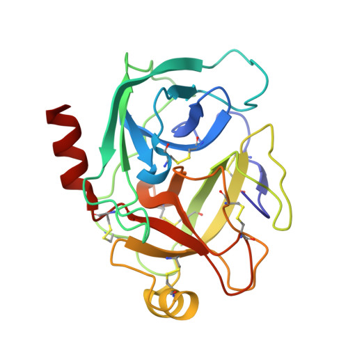
    New macrocyclic plasmin inhibitors based on our previously optimized P2-P3 core segment have been developed. In the first series, the P4 residue was modified, whereas the 4-amidinobenzylamide in P1 position was maintained. The originally used P4 benzylsulfonyl residue could be replaced by various sulfonyl- or urethane-like protecting groups. In the second series, the P1 benzamidine was modified and a strong potency and excellent selectivity was retained by incorporation of p-xylenediamine. Several analogues inhibit plasmin in the subnanomolar range, and their potency against related trypsin-like serine proteases including trypsin itself could be further reduced. Selected derivatives have been tested in a plasma fibrinolysis assay and are more effective than the reference inhibitor aprotinin. The crystal structure of one inhibitor was determined in complex with trypsin. The binding mode reveals a sterical clash of the inhibitor's linker segment with the 99-hairpin loop of trypsin, which is absent in plasmin.


  • Organizational Affiliation

    Department of Pharmacy, Institute of Pharmaceutical Chemistry, Philipps University Marburg , Marbacher Weg 6, D-35032 Marburg, Germany.


Macromolecules
Find similar proteins by:  (by identity cutoff)  |  3D Structure
Entity ID: 1
MoleculeChains Sequence LengthOrganismDetailsImage
Cationic trypsin223Bos taurusMutation(s): 0 
EC: 3.4.21.4
UniProt
Find proteins for P00760 (Bos taurus)
Explore P00760 
Go to UniProtKB:  P00760
Entity Groups  
Sequence Clusters30% Identity50% Identity70% Identity90% Identity95% Identity100% Identity
UniProt GroupP00760
Sequence Annotations
Expand
  • Reference Sequence
Small Molecules
Ligands 4 Unique
IDChains Name / Formula / InChI Key2D Diagram3D Interactions
5N7
Query on 5N7

Download Ideal Coordinates CCD File 
E [auth A](13S,16R)-N-(4-carbamimidoylbenzyl)-16-((N-cyclohexylsulfamoyl)amino)-3,9,15-trioxo-2,10,14-triaza-6(1,4)-piperazina-1, 11(1,4)-dibenzenacycloheptadecaphane-13-carboxamide
C42 H56 N10 O6 S
HGZHCKIUQIEHRY-PQQNNWGCSA-N
SO4
Query on SO4

Download Ideal Coordinates CCD File 
C [auth A]SULFATE ION
O4 S
QAOWNCQODCNURD-UHFFFAOYSA-L
ACT
Query on ACT

Download Ideal Coordinates CCD File 
D [auth A]ACETATE ION
C2 H3 O2
QTBSBXVTEAMEQO-UHFFFAOYSA-M
CA
Query on CA

Download Ideal Coordinates CCD File 
B [auth A]CALCIUM ION
Ca
BHPQYMZQTOCNFJ-UHFFFAOYSA-N
Binding Affinity Annotations 
IDSourceBinding Affinity
5N7 Binding MOAD:  5EG4 Ki: 21 (nM) from 1 assay(s)
Experimental Data & Validation

Experimental Data

  • Method: X-RAY DIFFRACTION
  • Resolution: 1.32 Å
  • R-Value Free: 0.218 
  • R-Value Work: 0.180 
  • R-Value Observed: 0.182 
  • Space Group: P 41 21 2
Unit Cell:
Length ( Å )Angle ( ˚ )
a = 61.502α = 90
b = 61.502β = 90
c = 102.239γ = 90
Software Package:
Software NamePurpose
PHENIXrefinement
HKL-2000data reduction
HKL-2000data scaling
PHASERphasing

Structure Validation

View Full Validation Report


Ligand Structure Quality Assessment 


Entry History 

Deposition Data

Revision History  (Full details and data files)

  • Version 1.0: 2016-07-13
    Type: Initial release
  • Version 1.1: 2016-07-27
    Changes: Database references
  • Version 1.2: 2024-01-10
    Changes: Data collection, Database references, Derived calculations, Refinement description, Structure summary