5EG2

SET7/9 N265A in complex with AdoHcy and TAF10 peptide


Experimental Data Snapshot

  • Method: X-RAY DIFFRACTION
  • Resolution: 1.55 Å
  • R-Value Free: 0.171 
  • R-Value Work: 0.147 
  • R-Value Observed: 0.148 

wwPDB Validation   3D Report Full Report


Ligand Structure Quality Assessment 


This is version 1.5 of the entry. See complete history


Literature

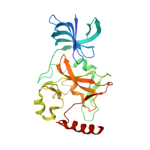
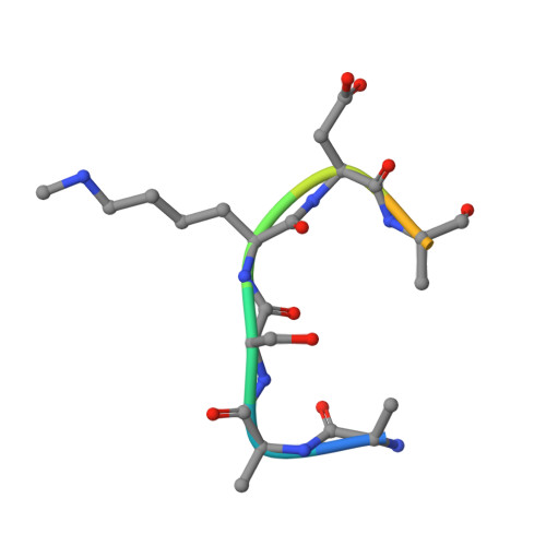
Sulfur-Oxygen Chalcogen Bonding Mediates AdoMet Recognition in the Lysine Methyltransferase SET7/9.

Fick, R.J.Kroner, G.M.Nepal, B.Magnani, R.Horowitz, S.Houtz, R.L.Scheiner, S.Trievel, R.C.

(2016) ACS Chem Biol 11: 748-754

  • DOI: https://doi.org/10.1021/acschembio.5b00852
  • Primary Citation of Related Structures:  
    5EG2

  • PubMed Abstract: 

    Recent studies have demonstrated that carbon-oxygen (CH···O) hydrogen bonds have important roles in S-adenosylmethionine (AdoMet) recognition and catalysis in methyltransferases. Here, we investigate noncovalent interactions that occur between the AdoMet sulfur cation and oxygen atoms in methyltransferase active sites. These interactions represent sulfur-oxygen (S···O) chalcogen bonds in which the oxygen atom donates a lone pair of electrons to the σ antibonding orbital of the AdoMet sulfur atom. Structural, biochemical, and computational analyses of an asparagine mutation in the lysine methyltransferase SET7/9 that abolishes AdoMet S···O chalcogen bonding reveal that this interaction enhances substrate binding affinity relative to the product S-adenosylhomocysteine. Corroborative quantum mechanical calculations demonstrate that sulfonium systems form strong S···O chalcogen bonds relative to their neutral thioether counterparts. An inspection of high-resolution crystal structures reveals the presence of AdoMet S···O chalcogen bonding in different classes of methyltransferases, illustrating that these interactions are not limited to SET domain methyltransferases. Together, these results demonstrate that S···O chalcogen bonds contribute to AdoMet recognition and can enable methyltransferases to distinguish between substrate and product.


  • Organizational Affiliation

    Department of Chemistry and Biochemistry, Utah State University , Logan, Utah 84322, United States.


Macromolecules
Find similar proteins by:  (by identity cutoff)  |  3D Structure
Entity ID: 1
MoleculeChains Sequence LengthOrganismDetailsImage
Histone-lysine N-methyltransferase SETD7262Homo sapiensMutation(s): 1 
Gene Names: SETD7KIAA1717KMT7SET7SET9
EC: 2.1.1.43
UniProt & NIH Common Fund Data Resources
Find proteins for Q8WTS6 (Homo sapiens)
Explore Q8WTS6 
Go to UniProtKB:  Q8WTS6
PHAROS:  Q8WTS6
GTEx:  ENSG00000145391 
Entity Groups  
Sequence Clusters30% Identity50% Identity70% Identity90% Identity95% Identity100% Identity
UniProt GroupQ8WTS6
Sequence Annotations
Expand
  • Reference Sequence

Find similar proteins by:  Sequence   |   3D Structure  

Entity ID: 2
MoleculeChains Sequence LengthOrganismDetailsImage
Transcription initiation factor TFIID subunit 1010Homo sapiensMutation(s): 0 
UniProt & NIH Common Fund Data Resources
Find proteins for Q12962 (Homo sapiens)
Explore Q12962 
Go to UniProtKB:  Q12962
PHAROS:  Q12962
GTEx:  ENSG00000166337 
Entity Groups  
Sequence Clusters30% Identity50% Identity70% Identity90% Identity95% Identity100% Identity
UniProt GroupQ12962
Sequence Annotations
Expand
  • Reference Sequence
Small Molecules
Ligands 2 Unique
IDChains Name / Formula / InChI Key2D Diagram3D Interactions
SAH
Query on SAH

Download Ideal Coordinates CCD File 
C [auth A]S-ADENOSYL-L-HOMOCYSTEINE
C14 H20 N6 O5 S
ZJUKTBDSGOFHSH-WFMPWKQPSA-N
NA
Query on NA

Download Ideal Coordinates CCD File 
D [auth A]SODIUM ION
Na
FKNQFGJONOIPTF-UHFFFAOYSA-N
Modified Residues  1 Unique
IDChains TypeFormula2D DiagramParent
MLZ
Query on MLZ
B
L-PEPTIDE LINKINGC7 H16 N2 O2LYS
Binding Affinity Annotations 
IDSourceBinding Affinity
SAH BindingDB:  5EG2 IC50: min: 3.00e+4, max: 4.70e+4 (nM) from 2 assay(s)
Experimental Data & Validation

Experimental Data

  • Method: X-RAY DIFFRACTION
  • Resolution: 1.55 Å
  • R-Value Free: 0.171 
  • R-Value Work: 0.147 
  • R-Value Observed: 0.148 
  • Space Group: P 32 2 1
Unit Cell:
Length ( Å )Angle ( ˚ )
a = 83.198α = 90
b = 83.198β = 90
c = 95.859γ = 120
Software Package:
Software NamePurpose
HKL-2000data reduction
HKL-2000data scaling
PHASERphasing
Cootmodel building
PHENIXrefinement
PDB_EXTRACTdata extraction

Structure Validation

View Full Validation Report


Ligand Structure Quality Assessment 


Entry History & Funding Information

Deposition Data


Funding OrganizationLocationGrant Number
National Science Foundation (NSF, United States)United StatesNSF-CHE-1508492

Revision History  (Full details and data files)

  • Version 1.0: 2016-01-13
    Type: Initial release
  • Version 1.1: 2016-02-03
    Changes: Database references
  • Version 1.2: 2016-03-30
    Changes: Database references
  • Version 1.3: 2017-09-20
    Changes: Author supporting evidence, Derived calculations
  • Version 1.4: 2019-11-27
    Changes: Author supporting evidence
  • Version 1.5: 2023-09-27
    Changes: Data collection, Database references, Derived calculations, Refinement description