5EEP

Crystal structure of E. coli CsdE


Experimental Data Snapshot

  • Method: X-RAY DIFFRACTION
  • Resolution: 2.40 Å
  • R-Value Free: 0.285 
  • R-Value Work: 0.229 
  • R-Value Observed: 0.231 

wwPDB Validation   3D Report Full Report


This is version 1.1 of the entry. See complete history


Literature

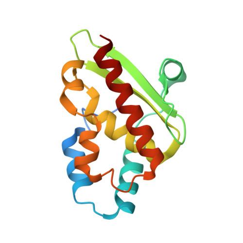
The crystal structure of Escherichia coli CsdE

Kenne, A.N.Kim, S.Park, S.Y.

(2016) Int J Biol Macromol 87: 317-321

  • DOI: https://doi.org/10.1016/j.ijbiomac.2016.02.071
  • Primary Citation of Related Structures:  
    5EEP

  • PubMed Abstract: 

    Sulfur incorporations both in the biosynthesis of sulfur-containing cofactors and in the sulfur-modifications of certain tRNAs are all mediated by the sulfur initially delivered from the cysteine desulfurases. Sulfur generated as persulfide from cysteine is transferred to the sulfur acceptor protein to further allow delivery to the required steps within an enzymatic process. CsdA which is one of the three cysteine desulfurases identified in Escherichia coli transfers sulfur to the non Fe-S sulfur-acceptor CsdE, however, the consequence of CsdE accepted sulfur is mostly unknown. In this study, we report the 2.4Å structure of free CsdE determined using X-ray crystallography, and compare the structure with the CsdE structure determined using NMR and also CsdE within the crystal CsdA-CsdE complex. Further analysis suggests that the positive electrostatic potential surfaces of CsdE may mediate interaction with a yet unidentified protein or possibly tRNA to deliver sulfur.


  • Organizational Affiliation

    School of Systems Biomedical Science, Soongsil University, Seoul 06978, Republic of Korea.


Macromolecules
Find similar proteins by:  (by identity cutoff)  |  3D Structure
Entity ID: 1
MoleculeChains Sequence LengthOrganismDetailsImage
CsdA-binding activator150Escherichia coliMutation(s): 0 
Gene Names: ECONIH1_16050SK78_01951
UniProt
Find proteins for P0AGF2 (Escherichia coli (strain K12))
Explore P0AGF2 
Go to UniProtKB:  P0AGF2
Entity Groups  
Sequence Clusters30% Identity50% Identity70% Identity90% Identity95% Identity100% Identity
UniProt GroupP0AGF2
Sequence Annotations
Expand
  • Reference Sequence
Experimental Data & Validation

Experimental Data

  • Method: X-RAY DIFFRACTION
  • Resolution: 2.40 Å
  • R-Value Free: 0.285 
  • R-Value Work: 0.229 
  • R-Value Observed: 0.231 
  • Space Group: P 43 21 2
Unit Cell:
Length ( Å )Angle ( ˚ )
a = 43.521α = 90
b = 43.521β = 90
c = 145.323γ = 90
Software Package:
Software NamePurpose
REFMACrefinement
HKL-2000data reduction
HKL-2000data scaling
HKL-2000phasing

Structure Validation

View Full Validation Report


Entry History 

Deposition Data

  • Released Date: 2016-06-08 
  • Deposition Author(s): Park, S.Y.

Revision History  (Full details and data files)

  • Version 1.0: 2016-06-08
    Type: Initial release
  • Version 1.1: 2024-03-20
    Changes: Data collection, Database references, Derived calculations