5E5Y | pdb_00005e5y

Quasi-racemic snakin-1 in P1 before radiation damage


Experimental Data Snapshot

  • Method: X-RAY DIFFRACTION
  • Resolution: 1.51 Å
  • R-Value Free: 
    0.226 (Depositor), 0.227 (DCC) 
  • R-Value Work: 
    0.179 (Depositor), 0.181 (DCC) 
  • R-Value Observed: 
    0.182 (Depositor) 

wwPDB Validation 3D Report Full Report

Validation slider image for 5E5Y

This is version 1.2 of the entry. See complete history

Literature

Radiation Damage and Racemic Protein Crystallography Reveal the Unique Structure of the GASA/Snakin Protein Superfamily.

Yeung, H.Squire, C.J.Yosaatmadja, Y.Panjikar, S.Lopez, G.Molina, A.Baker, E.N.Harris, P.W.Brimble, M.A.

(2016) Angew Chem Int Ed Engl 55: 7930-7933

  • DOI: https://doi.org/10.1002/anie.201602719
  • Primary Citation Related Structures: 
    5E5Q, 5E5T, 5E5Y

  • PubMed Abstract: 

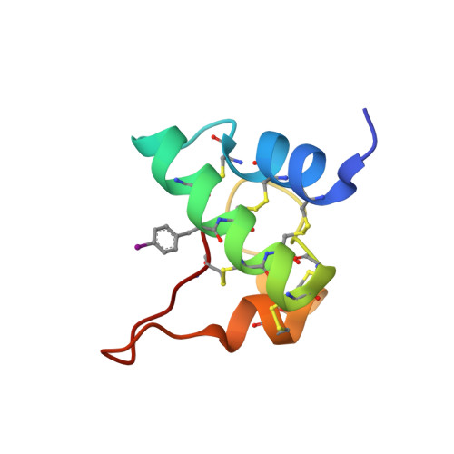
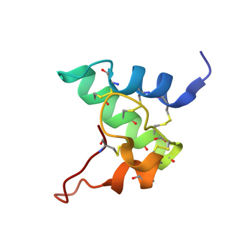
    Proteins from the GASA/snakin superfamily are common in plant proteomes and have diverse functions, including hormonal crosstalk, development, and defense. One 63-residue member of this family, snakin-1, an antimicrobial protein from potatoes, has previously been chemically synthesized in a fully active form. Herein the 1.5 Å structure of snakin-1, determined by a novel combination of racemic protein crystallization and radiation-damage-induced phasing (RIP), is reported. Racemic crystals of snakin-1 and quasi-racemic crystals incorporating an unnatural 4-iodophenylalanine residue were prepared from chemically synthesized d- and l-proteins. Breakage of the C-I bonds in the quasi-racemic crystals facilitated structure determination by RIP. The crystal structure reveals a unique protein fold with six disulfide crosslinks, presenting a distinct electrostatic surface that may target the protein to microbial cell surfaces.


  • Organizational Affiliation
    • School of Biological Sciences, The University of Auckland, 3A Symonds St, Auckland Central, 1010, New Zealand.

Macromolecule Content 

  • Total Structure Weight: 28.58 kDa 
  • Atom Count: 2,220 
  • Modeled Residue Count: 251 
  • Deposited Residue Count: 252 
  • Unique protein chains: 2

Macromolecules

Find similar proteins by:|  3D Structure
Entity ID: 1
MoleculeChains  Sequence LengthOrganismDetailsImage
Snakin-1
A, C
63Solanum tuberosumMutation(s): 1 
UniProt
Find proteins for Q948Z4 (Solanum tuberosum)
Explore Q948Z4 
Go to UniProtKB:  Q948Z4
Entity Groups
Sequence Clusters30% Identity50% Identity70% Identity90% Identity95% Identity100% Identity
UniProt GroupQ948Z4
Sequence Annotations
Expand
Reference Sequence
Find similar proteins by:|  3D Structure
Entity ID: 2
MoleculeChains  Sequence LengthOrganismDetailsImage
D- snakin-1
B, D
63Solanum tuberosumMutation(s): 0 
UniProt
Find proteins for Q948Z4 (Solanum tuberosum)
Explore Q948Z4 
Go to UniProtKB:  Q948Z4
Entity Groups
Sequence Clusters30% Identity50% Identity70% Identity90% Identity95% Identity100% Identity
UniProt GroupQ948Z4
Sequence Annotations
Expand
Reference Sequence

Small Molecules

Ligands 2 Unique
IDChains Name / Formula / InChI Key2D Diagram3D Interactions
EDO

Query on EDO


Download:Ideal Coordinates CCD File
E [auth A],
F [auth A],
H [auth B],
K [auth C],
O [auth D]
1,2-ETHANEDIOL
C2 H6 O2
LYCAIKOWRPUZTN-UHFFFAOYSA-N
FMT

Query on FMT


Download:Ideal Coordinates CCD File
G [auth B]
I [auth C]
J [auth C]
L [auth D]
M [auth D]
G [auth B],
I [auth C],
J [auth C],
L [auth D],
M [auth D],
N [auth D]
FORMIC ACID
C H2 O2
BDAGIHXWWSANSR-UHFFFAOYSA-N
Modified Residues  1 Unique
IDChains TypeFormula2D DiagramParent
PHI
Query on PHI
A, C
L-PEPTIDE LINKINGC9 H10 I N O2PHE
DAL
Query on DAL
B, D
D-PEPTIDE LINKINGC3 H7 N O2

--

DAR
Query on DAR
B, D
D-PEPTIDE LINKINGC6 H15 N4 O2

--

DAS
Query on DAS
B, D
D-PEPTIDE LINKINGC4 H7 N O4

--

DCY
Query on DCY
B, D
D-PEPTIDE LINKINGC3 H7 N O2 S

--

DGL
Query on DGL
B, D
D-PEPTIDE LINKINGC5 H9 N O4

--

DHI
Query on DHI
B, D
D-PEPTIDE LINKINGC6 H10 N3 O2

--

DIL
Query on DIL
B, D
D-PEPTIDE LINKINGC6 H13 N O2

--

DLE
Query on DLE
B, D
D-PEPTIDE LINKINGC6 H13 N O2

--

DLY
Query on DLY
B, D
D-PEPTIDE LINKINGC6 H14 N2 O2

--

DPN
Query on DPN
B, D
D-PEPTIDE LINKINGC9 H11 N O2

--

DPR
Query on DPR
B, D
D-PEPTIDE LINKINGC5 H9 N O2

--

DSG
Query on DSG
B, D
D-PEPTIDE LINKINGC4 H8 N2 O3

--

DSN
Query on DSN
B, D
D-PEPTIDE LINKINGC3 H7 N O3

--

DTH
Query on DTH
B, D
D-PEPTIDE LINKINGC4 H9 N O3

--

DTY
Query on DTY
B, D
D-PEPTIDE LINKINGC9 H11 N O3

--

DVA
Query on DVA
B, D
D-PEPTIDE LINKINGC5 H11 N O2

--

Experimental Data & Validation

Experimental Data

  • Method: X-RAY DIFFRACTION
  • Resolution: 1.51 Å
  • R-Value Free:  0.226 (Depositor), 0.227 (DCC) 
  • R-Value Work:  0.179 (Depositor), 0.181 (DCC) 
  • R-Value Observed: 0.182 (Depositor) 
Space Group: P 1
Unit Cell:
Length ( Å )Angle ( ˚ )
a = 31.233α = 93
b = 37.415β = 90.58
c = 50.303γ = 102.57
Software Package:
Software NamePurpose
PHENIXrefinement
Aimlessdata scaling
XDSdata reduction
SHELXDEphasing

Structure Validation

View Full Validation Report


Entry History 

& Funding Information

Deposition Data


Funding OrganizationLocationGrant Number
Royal Society of New ZealandNew ZealandUOA1115

Revision History  (Full details and data files)

  • Version 1.0: 2016-05-18
    Type: Initial release
  • Version 1.1: 2016-07-20
    Changes: Database references
  • Version 1.2: 2025-04-02
    Changes: Data collection, Database references, Derived calculations, Structure summary