5DZT

Crystal structure of class II lanthipeptide synthetase CylM in complex with AMP


Experimental Data Snapshot

  • Method: X-RAY DIFFRACTION
  • Resolution: 2.20 Å
  • R-Value Free: 0.275 
  • R-Value Work: 0.244 
  • R-Value Observed: 0.245 

wwPDB Validation   3D Report Full Report


Ligand Structure Quality Assessment 


This is version 1.3 of the entry. See complete history


Literature

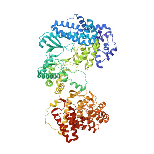
The enterococcal cytolysin synthetase has an unanticipated lipid kinase fold.

Dong, S.H.Tang, W.Lukk, T.Yu, Y.Nair, S.K.van der Donk, W.A.

(2015) Elife 4

  • DOI: https://doi.org/10.7554/eLife.07607
  • Primary Citation of Related Structures:  
    5DZT

  • PubMed Abstract: 

    The enterococcal cytolysin is a virulence factor consisting of two post-translationally modified peptides that synergistically kill human immune cells. Both peptides are made by CylM, a member of the LanM lanthipeptide synthetases. CylM catalyzes seven dehydrations of Ser and Thr residues and three cyclization reactions during the biosynthesis of the cytolysin large subunit. We present here the 2.2 Å resolution structure of CylM, the first structural information on a LanM. Unexpectedly, the structure reveals that the dehydratase domain of CylM resembles the catalytic core of eukaryotic lipid kinases, despite the absence of clear sequence homology. The kinase and phosphate elimination active sites that affect net dehydration are immediately adjacent to each other. Characterization of mutants provided insights into the mechanism of the dehydration process. The structure is also of interest because of the interactions of human homologs of lanthipeptide cyclases with kinases such as mammalian target of rapamycin.


  • Organizational Affiliation

    Department of Biochemistry, University of Illinois at Urbana-Champaign, Urbana, United States.


Macromolecules
Find similar proteins by:  (by identity cutoff)  |  3D Structure
Entity ID: 1
MoleculeChains Sequence LengthOrganismDetailsImage
CylM993Enterococcus faecalisMutation(s): 0 
Gene Names: ef0046cylM
UniProt
Find proteins for Q8KUA3 (Enterococcus faecalis)
Explore Q8KUA3 
Go to UniProtKB:  Q8KUA3
Entity Groups  
Sequence Clusters30% Identity50% Identity70% Identity90% Identity95% Identity100% Identity
UniProt GroupQ8KUA3
Sequence Annotations
Expand
  • Reference Sequence
Small Molecules
Ligands 2 Unique
IDChains Name / Formula / InChI Key2D Diagram3D Interactions
AMP
Query on AMP

Download Ideal Coordinates CCD File 
C [auth A]ADENOSINE MONOPHOSPHATE
C10 H14 N5 O7 P
UDMBCSSLTHHNCD-KQYNXXCUSA-N
ZN
Query on ZN

Download Ideal Coordinates CCD File 
B [auth A]ZINC ION
Zn
PTFCDOFLOPIGGS-UHFFFAOYSA-N
Experimental Data & Validation

Experimental Data

  • Method: X-RAY DIFFRACTION
  • Resolution: 2.20 Å
  • R-Value Free: 0.275 
  • R-Value Work: 0.244 
  • R-Value Observed: 0.245 
  • Space Group: P 21 21 21
Unit Cell:
Length ( Å )Angle ( ˚ )
a = 51.191α = 90
b = 90.699β = 90
c = 246.361γ = 90
Software Package:
Software NamePurpose
REFMACrefinement
Cootmodel building
SHELXDphasing
SHARPphasing
HKL-2000data scaling
HKL-2000data reduction

Structure Validation

View Full Validation Report


Ligand Structure Quality Assessment 


Entry History & Funding Information

Deposition Data


Funding OrganizationLocationGrant Number
National Institutes of Health/National Institute of General Medical Sciences (NIH/NIGMS)United StatesRO1 GM079038

Revision History  (Full details and data files)

  • Version 1.0: 2015-10-14
    Type: Initial release
  • Version 1.1: 2017-09-27
    Changes: Author supporting evidence, Derived calculations, Refinement description
  • Version 1.2: 2019-12-25
    Changes: Author supporting evidence
  • Version 1.3: 2024-03-06
    Changes: Data collection, Database references