5DQ0 | pdb_00005dq0

Structure of human neuropilin-2 b1 domain with novel and unique zinc binding site


Experimental Data Snapshot

  • Method: X-RAY DIFFRACTION
  • Resolution: 1.80 Å
  • R-Value Free: 
    0.217 (Depositor), 0.226 (DCC) 
  • R-Value Work: 
    0.177 (Depositor), 0.188 (DCC) 
  • R-Value Observed: 
    0.179 (Depositor) 

Starting Model: experimental
View more details

wwPDB Validation 3D Report Full Report

Validation slider image for 5DQ0

This is version 1.3 of the entry. See complete history

Literature

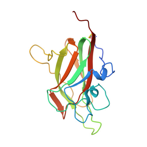
Structural studies of neuropilin-2 reveal a zinc ion binding site remote from the vascular endothelial growth factor binding pocket.

Tsai, Y.C.Fotinou, C.Rana, R.Yelland, T.Frankel, P.Zachary, I.Djordjevic, S.

(2016) FEBS J 283: 1921-1934

  • DOI: https://doi.org/10.1111/febs.13711
  • Primary Citation Related Structures: 
    5DN2, 5DQ0

  • PubMed Abstract: 

    Neuropilin-2 is a transmembrane receptor involved in lymphangiogenesis and neuronal development. In adults, neuropilin-2 and its homologous protein neuropilin-1 have been implicated in cancers and infection. Molecular determinants of the ligand selectivity of neuropilins are poorly understood. We have identified and structurally characterized a zinc ion binding site on human neuropilin-2. The neuropilin-2-specific zinc ion binding site is located near the interface between domains b1 and b2 in the ectopic region of the protein, remote from the neuropilin binding site for its physiological ligand, i.e. vascular endothelial growth factor. We also present an X-ray crystal structure of the neuropilin-2 b1 domain in a complex with the C-terminal sub-domain of VEGF-A. Zn(2+) binding to neuropilin-2 destabilizes the protein structure but this effect was counteracted by heparin, suggesting that modifications by glycans and zinc in the extracellular matrix may affect functional neuropilin-2 ligand binding and signalling activity.


  • Organizational Affiliation
    • Institute of Structural and Molecular Biology, University College London, London, UK.

Macromolecule Content 

  • Total Structure Weight: 18.96 kDa 
  • Atom Count: 1,467 
  • Modeled Residue Count: 157 
  • Deposited Residue Count: 162 
  • Unique protein chains: 1

Macromolecules

Find similar proteins by:|  3D Structure
Entity ID: 1
MoleculeChains  Sequence LengthOrganismDetailsImage
Neuropilin-2162Homo sapiensMutation(s): 0 
Gene Names: NRP2VEGF165R2
UniProt & NIH Common Fund Data Resources
Find proteins for O60462 (Homo sapiens)
Explore O60462 
Go to UniProtKB:  O60462
PHAROS:  O60462
GTEx:  ENSG00000118257 
Entity Groups
Sequence Clusters30% Identity50% Identity70% Identity90% Identity95% Identity100% Identity
UniProt GroupO60462
Sequence Annotations
Expand
Reference Sequence

Small Molecules

Ligands 4 Unique
IDChains Name / Formula / InChI Key2D Diagram3D Interactions
MES

Query on MES


Download:Ideal Coordinates CCD File
I [auth A]2-(N-MORPHOLINO)-ETHANESULFONIC ACID
C6 H13 N O4 S
SXGZJKUKBWWHRA-UHFFFAOYSA-N
ZN

Query on ZN


Download:Ideal Coordinates CCD File
B [auth A],
C [auth A],
D [auth A]
ZINC ION
Zn
PTFCDOFLOPIGGS-UHFFFAOYSA-N
EDO

Query on EDO


Download:Ideal Coordinates CCD File
G [auth A],
H [auth A]
1,2-ETHANEDIOL
C2 H6 O2
LYCAIKOWRPUZTN-UHFFFAOYSA-N
CL

Query on CL


Download:Ideal Coordinates CCD File
E [auth A],
F [auth A]
CHLORIDE ION
Cl
VEXZGXHMUGYJMC-UHFFFAOYSA-M

Experimental Data & Validation

Experimental Data

  • Method: X-RAY DIFFRACTION
  • Resolution: 1.80 Å
  • R-Value Free:  0.217 (Depositor), 0.226 (DCC) 
  • R-Value Work:  0.177 (Depositor), 0.188 (DCC) 
  • R-Value Observed: 0.179 (Depositor) 
Space Group: C 2 2 21
Unit Cell:
Length ( Å )Angle ( ˚ )
a = 99.39α = 90
b = 105.27β = 90
c = 45.75γ = 90
Software Package:
Software NamePurpose
REFMACrefinement
Cootmodel building
SCALAdata scaling
MOSFLMdata reduction
PHASERphasing

Structure Validation

View Full Validation Report


Entry History 

& Funding Information

Deposition Data


Funding OrganizationLocationGrant Number
British Heart FoundationUnited KingdomPG/10/52/28448

Revision History  (Full details and data files)

  • Version 1.0: 2016-09-28
    Type: Initial release
  • Version 1.1: 2024-01-10
    Changes: Data collection, Database references, Derived calculations, Refinement description
  • Version 1.2: 2024-10-23
    Changes: Structure summary
  • Version 1.3: 2026-03-25
    Changes: Database references