Structure and Mutational Analyses of Escherichia coli ZapD Reveal Charged Residues Involved in FtsZ Filament Bundling.
Roach, E.J., Wroblewski, C., Seidel, L., Berezuk, A.M., Brewer, D., Kimber, M.S., Khursigara, C.M.(2016) J Bacteriol 198: 1683-1693
- PubMed: 27021560 Search on PubMedSearch on PubMed Central
- DOI: https://doi.org/10.1128/JB.00969-15
- Primary Citation Related Structures:
5DKO - PubMed Abstract:
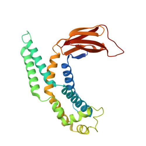
Bacterial cell division is an essential and highly coordinated process. It requires the polymerization of the tubulin homologue FtsZ to form a dynamic ring (Z-ring) at midcell. Z-ring formation relies on a group of FtsZ-associated proteins (Zap) for stability throughout the process of division. In Escherichia coli, there are currently five Zap proteins (ZapA through ZapE), of which four (ZapA, ZapB, ZapC, and ZapD) are small soluble proteins that act to bind and bundle FtsZ filaments. In particular, ZapD forms a functional dimer and interacts with the C-terminal tail of FtsZ, but little is known about its structure and mechanism of action. Here, we present the crystal structure of Escherichia coli ZapD and show it forms a symmetrical dimer with centrally located α-helices flanked by β-sheet domains. Based on the structure of ZapD and its chemical cross-linking to FtsZ, we targeted nine charged ZapD residues for modification by site-directed mutagenesis. Using in vitro FtsZ sedimentation assays, we show that residues R56, R221, and R225 are important for bundling FtsZ filaments, while transmission electron microscopy revealed that altering these residues results in different FtsZ bundle morphology compared to those of filaments bundled with wild-type ZapD. ZapD residue R116 also showed altered FtsZ bundle morphology but levels of FtsZ bundling similar to that of wild-type ZapD. Together, these results reveal that ZapD residues R116, R221, and R225 likely participate in forming a positively charged binding pocket that is critical for bundling FtsZ filaments. Z-ring assembly underpins the formation of the essential cell division complex known as the divisome and is required for recruitment of downstream cell division proteins. ZapD is one of several proteins in E. coli that associates with the Z-ring to promote FtsZ bundling and aids in the overall fitness of the division process. In the present study, we describe the dimeric structure of E. coli ZapD and identify residues that are critical for FtsZ bundling. Together, these results advance our understanding about the formation and dynamics of the Z-ring prior to bacterial cell division.
- Department of Molecular and Cellular Biology, University of Guelph, Guelph, Ontario, Canada.
Organizational Affiliation: