5D4K | pdb_00005d4k

Crystal structure of the human polymeric Ig receptor (pIgR) ectodomain

  • Classification: IMMUNE SYSTEM
  • Organism(s): Homo sapiens
  • Expression System: Homo sapiens
  • Mutation(s): Yes 

  • Deposited: 2015-08-07 Released: 2016-03-16 
  • Deposition Author(s): Stadtmueller, B.M., Bjorkman, P.J.
  • Funding Organization(s): National Institutes of Health/National Institute Of Allergy and Infectious Diseases (NIH/NIAID), Cancer Research Institute

Experimental Data Snapshot

  • Method: X-RAY DIFFRACTION
  • Resolution: 2.60 Å
  • R-Value Free: 
    0.254 (Depositor), 0.244 (DCC) 
  • R-Value Work: 
    0.201 (Depositor) 
  • R-Value Observed: 
    0.203 (Depositor) 

Starting Model: experimental
View more details

wwPDB Validation 3D Report Full Report

Validation slider image for 5D4K

Ligand Structure Quality Assessment 


This is version 2.2 of the entry. See complete history

Literature

The structure and dynamics of secretory component and its interactions with polymeric immunoglobulins.

Stadtmueller, B.M.Huey-Tubman, K.E.Lopez, C.J.Yang, Z.Hubbell, W.L.Bjorkman, P.J.

(2016) Elife 5

  • DOI: https://doi.org/10.7554/eLife.10640
  • Primary Citation Related Structures: 
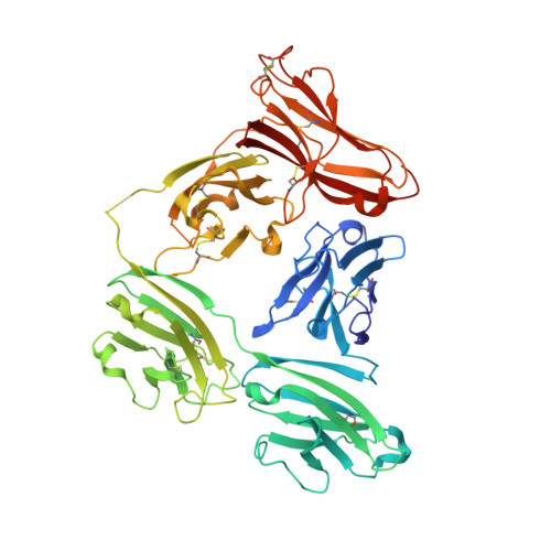
    5D4K, 5F1S

  • PubMed Abstract: 

    As a first-line vertebrate immune defense, the polymeric immunoglobulin receptor (pIgR) transports polymeric IgA and IgM across epithelia to mucosal secretions, where the cleaved ectodomain (secretory component; SC) becomes a component of secretory antibodies, or when unliganded, binds and excludes bacteria. Here we report the 2.6Å crystal structure of unliganded human SC (hSC) and comparisons with a 1.7Å structure of teleost fish SC (tSC), an early pIgR ancestor. The hSC structure comprises five immunoglobulin-like domains (D1-D5) arranged as a triangle, with an interface between ligand-binding domains D1 and D5. Electron paramagnetic resonance measurements confirmed the D1-D5 interface in solution and revealed that it breaks upon ligand binding. Together with binding studies of mutant and chimeric SCs, which revealed domain contributions to secretory antibody formation, these results provide detailed models for SC structure, address pIgR evolution, and demonstrate that SC uses multiple conformations to protect mammals from pathogens.


  • Organizational Affiliation
    • Division of Biology and Biological Engineering, California Institute of Technology, Pasadena, United States.

Macromolecule Content 

  • Total Structure Weight: 125.41 kDa 
  • Atom Count: 8,530 
  • Modeled Residue Count: 1,075 
  • Deposited Residue Count: 1,114 
  • Unique protein chains: 1

Macromolecules

Find similar proteins by:|  3D Structure
Entity ID: 1
MoleculeChains  Sequence LengthOrganismDetailsImage
Polymeric immunoglobulin receptor
A, B
557Homo sapiensMutation(s): 1 
Gene Names: PIGR
UniProt & NIH Common Fund Data Resources
Find proteins for P01833 (Homo sapiens)
Explore P01833 
Go to UniProtKB:  P01833
PHAROS:  P01833
GTEx:  ENSG00000162896 
Entity Groups
Sequence Clusters30% Identity50% Identity70% Identity90% Identity95% Identity100% Identity
UniProt GroupP01833
Glycosylation
Glycosylation Sites: 4Go to GlyGen: P01833-1
Sequence Annotations
Expand
Reference Sequence

Oligosaccharides

Help  
Entity ID: 2
MoleculeChains Length2D Diagram GlycosylationD Interactions
alpha-L-fucopyranose-(1-6)-2-acetamido-2-deoxy-beta-D-glucopyranose
C, D
2N-Glycosylation
Glycosylation Resources
GlyTouCan: G86851RC
GlyCosmos: G86851RC
GlyGen: G86851RC

Small Molecules

Ligands 2 Unique
IDChains Name / Formula / InChI Key2D Diagram3D Interactions
NAG

Query on NAG


Download:Ideal Coordinates CCD File
E [auth A]
F [auth A]
G [auth A]
J [auth B]
K [auth B]
E [auth A],
F [auth A],
G [auth A],
J [auth B],
K [auth B],
L [auth B]
2-acetamido-2-deoxy-beta-D-glucopyranose
C8 H15 N O6
OVRNDRQMDRJTHS-FMDGEEDCSA-N
SO4

Query on SO4


Download:Ideal Coordinates CCD File
H [auth A],
I [auth A],
M [auth B],
N [auth B]
SULFATE ION
O4 S
QAOWNCQODCNURD-UHFFFAOYSA-L

Experimental Data & Validation

Experimental Data

  • Method: X-RAY DIFFRACTION
  • Resolution: 2.60 Å
  • R-Value Free:  0.254 (Depositor), 0.244 (DCC) 
  • R-Value Work:  0.201 (Depositor) 
  • R-Value Observed: 0.203 (Depositor) 
Space Group: P 1 21 1
Unit Cell:
Length ( Å )Angle ( ˚ )
a = 61.258α = 90
b = 242.434β = 114.89
c = 63.046γ = 90
Software Package:
Software NamePurpose
PHENIXrefinement
SCALAdata scaling
PHASERphasing
PDB_EXTRACTdata extraction
Cootmodel building
Blu-Icedata collection
XDSdata reduction

Structure Validation

View Full Validation Report


Ligand Structure Quality Assessment 


Entry History 

& Funding Information

Deposition Data


Funding OrganizationLocationGrant Number
National Institutes of Health/National Institute Of Allergy and Infectious Diseases (NIH/NIAID)United StatesR01AI04123
Cancer Research InstituteUnited StatesIrving Postdoctoral Fellowship

Revision History  (Full details and data files)

  • Version 1.0: 2016-03-16
    Type: Initial release
  • Version 1.1: 2017-09-27
    Changes: Author supporting evidence, Derived calculations
  • Version 1.2: 2019-12-11
    Changes: Author supporting evidence, Data collection
  • Version 2.0: 2020-07-29
    Type: Remediation
    Reason: Carbohydrate remediation
    Changes: Atomic model, Data collection, Derived calculations, Structure summary
  • Version 2.1: 2023-09-27
    Changes: Data collection, Database references, Derived calculations, Refinement description, Structure summary
  • Version 2.2: 2024-11-13
    Changes: Structure summary