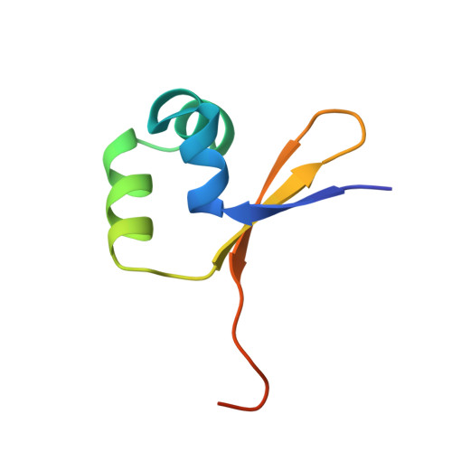
5CRO

REFINED STRUCTURE OF CRO REPRESSOR PROTEIN FROM BACTERIOPHAGE LAMBDA


Experimental Data Snapshot

  • Method: X-RAY DIFFRACTION
  • Resolution: 2.30 Å
  • R-Value Work: 0.193 

wwPDB Validation   3D Report Full Report


This is version 1.2 of the entry. See complete history


Literature

Refined structure of Cro repressor protein from bacteriophage lambda suggests both flexibility and plasticity.

Ohlendorf, D.H.Tronrud, D.E.Matthews, B.W.

(1998) J Mol Biol 280: 129-136

  • DOI: https://doi.org/10.1006/jmbi.1998.1849
  • Primary Citation of Related Structures:  
    5CRO

  • PubMed Abstract: 
  • The structure of the Cro repressor protein from phage lambda has been refined to a crystallographic R-value of 19.3% at 2.3 A resolution. The re fined model supports the structure as originally described in 1981 and provides a basis for comparison with the Cro-operator complex described in the accompanying paper ...

    The structure of the Cro repressor protein from phage lambda has been refined to a crystallographic R-value of 19.3% at 2.3 A resolution. The re fined model supports the structure as originally described in 1981 and provides a basis for comparison with the Cro-operator complex described in the accompanying paper. Changes in structure seen in different crystal forms and modifications of Cro suggest that the individual subunits are somewhat plastic in nature. In addition, the dimer of Cro suggests a high degree of flexibility, which may be important in forming the Cro-DNA complex. The structure of the Cro subunit as determined by NMR agrees reasonably well with that in the crystals (root-mean-square discrepancy of about 2 A for all atoms). There are, however, only a limited number of intersubunit distance constraints and, presumably for this reason, the different NMR models for the dimer vary substantially among themselves (discrepancies of 1.3 to 5.5 A). Because of this variation it is not possible to say whether the range of discrepancies between the X-ray and NMR Cro dimers (2.9 to 7.5 A) represent a significant difference between the X-ray and solution structures. It has previously been proposed that substitutions of Tyr26 in Cro increase thermal stability by the "reverse hydrophobic effect", i.e. by exposing 40% more hydrophobic surface to solvent in the folded form than in the unfolded state. The refined structure, however, suggests that Tyr26 is equally solvent exposed in the folded and unfolded states. The most stabilizing substitution is Tyr26-->Asp and in this case it appears that interaction with an alpha-helix dipole is at least partly responsible for the enhanced stability.


    Related Citations: 
    • Different Interactions Used by Cro Repressor in Specific and Nonspecific DNA Binding
      Takeda, Y., Kim, J.G., Caday, C.G., Steers Junior, E., Ohlendorf, D.H., Anderson, W.F., Matthews, B.W.
      (1986) J Biol Chem 261: 8608
    • Use of Protein Sequence and Structure to Infer Distant Evolutionary Relationships
      Brennan, R.G., Weaver, L.H., Matthews, B.W.
      (1986) Chem Scr 26B: 251
    • The Structure of Cro Repressor Protein
      Takeda, Y., Ohlendorf, D.H., Anderson, W.F., Matthews, B.W.
      (1985) Biological Macromolecules And Assemblies V 2: Nucleic Acids And Interactive Proteins 2: 234
    • High Resolution Structural Studies of Cro Repressor Protein and Implications for DNA Recognition
      Ohlendorf, D.H., Anderson, W.F., Takeda, Y., Matthews, B.W.
      (1983) J Biomol Struct Dyn 1: 553
    • DNA-Binding Proteins
      Takeda, Y., Ohlendorf, D.H., Anderson, W.F., Matthews, B.W.
      (1983) Science 221: 1020
    • Comparison of the Structures of Cro and Lambda Repressor Proteins from Bacteriophage Lambda
      Ohlendorf, D.H., Anderson, W.F., Lewis, M., Pabo, C.O., Matthews, B.W.
      (1983) J Mol Biol 169: 757
    • Crystallographic Data for Complexes of the Cro Repressor with DNA
      Anderson, W.F., Cygler, M., Vandonselaar, M., Ohlendorf, D.H., Matthews, B.W., Kim, J., Takeda, Y.
      (1983) J Mol Biol 168: 903
    • Structural Studies of Protein-Nucleic Acid Interactions
      Ohlendorf, D.H., Matthews, B.W.
      (1983) Annu Rev Biophys Bioeng 12: 259
    • How Does Cro Repressor Recognize its DNA Target Sites?
      Matthews, B.W., Ohlendorf, D.H., Anderson, W.F., Fisher, R.G., Takeda, Y.
      (1983) Trends Biochem Sci 8: 25
    • Cro Repressor Protein and its Interaction with DNA
      Matthews, B.W., Ohlendorf, D.H., Anderson, W.F., Fisher, R.G., Takeda, Y.
      (1983) Cold Spring Harb Symp Quant Biol 47: 427
    • Many Gene-Regulatory Proteins Appear to Have a Similar Alpha-Helical Fold that Binds DNA and Evolved from a Common Precursor
      Ohlendorf, D.H., Anderson, W.F., Matthews, B.W.
      (1983) J Mol Evol 19: 109
    • The Molecular Basis of DNA-Protein Recognition Inferred from the Structure of Cro Repressor
      Ohlendorf, D.H., Anderson, W.F., Fisher, R.G., Takeda, Y., Matthews, B.W.
      (1982) Nature 298: 718
    • Structural Similarity in the DNA-Binding Domains of Catabolite Gene Activator and Cro Repressor Proteins
      Steitz, T.A., Ohlendorf, D.H., Mckay, D.B., Anderson, W.F., Matthews, B.W.
      (1982) Proc Natl Acad Sci U S A 79: 3097
    • Structure of the DNA-Binding Region of Lac Repressor Inferred from its Homology with Cro Repressor
      Matthews, B.W., Ohlendorf, D.H., Anderson, W.F., Takeda, Y.
      (1982) Proc Natl Acad Sci U S A 79: 1428
    • Proposed Alpha-Helical Super-Secondary Structure Associated with Protein-DNA Recognition
      Anderson, W.F., Takeda, Y., Ohlendorf, D.H., Matthews, B.W.
      (1982) J Mol Biol 159: 745
    • Structure of the Cro Repressor from Bacteriophage Lambda and its Interaction with DNA
      Anderson, W.F., Ohlendorf, D.H., Takeda, Y., Matthews, B.W.
      (1981) Nature 290: 754
    • The Structure of a Repressor. Crystallographic Data for the Cro Regulatory Protein of Bacteriophage Lambda
      Anderson, W.F., Matthews, B.W., Takeda, Y., Echols, H.
      (1979) J Mol Biol 130: 507
    • Amino Acid Sequence of Cro Regulatory Protein of Bacteriophage Lambda
      Hsiang, M.W., Cole, R.D., Takeda, Y., Echols, H.
      (1977) Nature 270: 275

    Organizational Affiliation

    Institute of Molecular Biology Howard Hughes Medical Institute and Department of Physics, University of Oregon, Eugene, OR, 97403-1229, USA.


Macromolecules
Find similar proteins by:  (by identity cutoff)  |  3D Structure
Entity ID: 1
MoleculeChainsSequence LengthOrganismDetailsImage
CRO REPRESSOR PROTEINA [auth O],
B [auth A],
C [auth B],
D [auth C]
66Lambdavirus lambdaMutation(s): 0 
Gene Names: crolambdap57
UniProt
Find proteins for P03040 (Escherichia phage lambda)
Explore P03040 
Go to UniProtKB:  P03040
Entity Groups  
Sequence Clusters30% Identity50% Identity70% Identity90% Identity95% Identity100% Identity
UniProt GroupP03040
Protein Feature View
Expand
  • Reference Sequence
Small Molecules
Ligands 1 Unique
IDChainsName / Formula / InChI Key2D Diagram3D Interactions
PO4
Query on PO4

Download Ideal Coordinates CCD File 
E [auth O],
F [auth O]
PHOSPHATE ION
O4 P
NBIIXXVUZAFLBC-UHFFFAOYSA-K
 Ligand Interaction
Experimental Data & Validation

Experimental Data

  • Method: X-RAY DIFFRACTION
  • Resolution: 2.30 Å
  • R-Value Work: 0.193 
  • Space Group: H 3 2
Unit Cell:
Length ( Å )Angle ( ˚ )
a = 91.6α = 90
b = 91.6β = 90
c = 268.5γ = 120
Software Package:
Software NamePurpose
LYNNmodel building
TNTrefinement
OSCTSTdata reduction
VENUSdata reduction
CCP4data scaling
ODPROCdata scaling
NEWREFphasing

Structure Validation

View Full Validation Report


Entry History 

Deposition Data

Revision History  (Full details and data files)

  • Version 1.0: 1998-06-17
    Type: Initial release
  • Version 1.1: 2008-03-25
    Changes: Version format compliance
  • Version 1.2: 2011-07-13
    Changes: Version format compliance