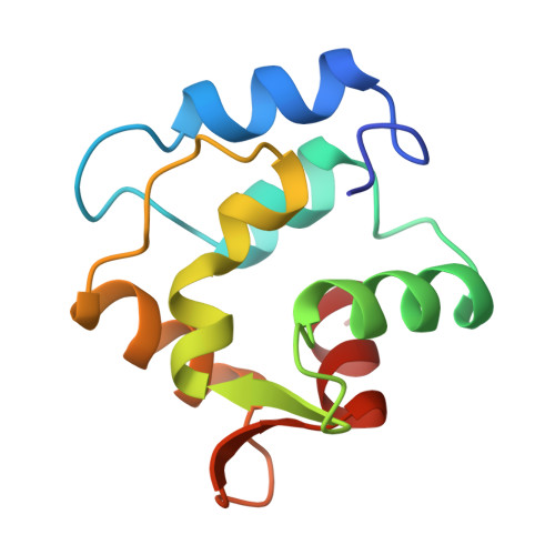
RESTRAINED LEAST SQUARES REFINEMENT OF NATIVE (CALCIUM) AND CADMIUM-SUBSTITUTED CARP PARVALBUMIN USING X-RAY CRYSTALLOGRAPHIC DATA AT 1.6-ANGSTROMS RESOLUTION
Restrained least squares refinement of native (calcium) and cadmium-substituted carp parvalbumin using X-ray crystallographic data at 1.6-A resolution.
Primary Citation of Related Structures:   1CDP, 5CPV
PubMed Abstract: 
Carp parvalbumin coordinates calcium through one carbonyl oxygen atom and the oxygen-containing side chains of 5 amino acid residues, or 4 residues and a water molecule, in a helix-loop-helix structural motif. Other calcium-binding proteins, including calmodulin and troponin C, also possess this unique calcium-binding design, which is designated EF-hand or calmodulin fold ...
Carp parvalbumin coordinates calcium through one carbonyl oxygen atom and the oxygen-containing side chains of 5 amino acid residues, or 4 residues and a water molecule, in a helix-loop-helix structural motif. Other calcium-binding proteins, including calmodulin and troponin C, also possess this unique calcium-binding design, which is designated EF-hand or calmodulin fold. Parvalbumin has two such sites, labeled CD and EF. Each of the calcium-binding sites of refined structures of proteins belonging to this group has a 7-oxygen coordination sphere except those of the structure of parvalbumin as it was reported in 1975. This structure had been refined at 1.9 A using difference Fourier techniques on film data. The CD site appeared to be 6-coordinate and the EF site 8-coordinate. Results of NMR experiments using 113Cd-substituted parvalbumin, however, indicate that the sites are similar to one another with coordination number greater than 6. To resolve the inconsistency between crystallographic and NMR results, 1.6 A area detector data was collected for native and cadmium-substituted parvalbumin; the structures have been refined to R factors of 18.7% and 16.4%, respectively, with acceptable geometry and low errors in atomic coordinates. Differences between the parvalbumin structure described in 1975 and the present structure are addressed, including the discovery of 7-coordination for both the CD and EF sites.
Related Citations: 
The Coordination Polyhedron of Ca2+,Cd2+ in Parvalbumin Swain, A.L., Amma, E.L. (1989) Inorganica Chim Acta 163: 5
Refinement of the Structure of Carp Muscle Calcium-Binding Parvalbumin by Model Building and Difference Fourier Analysis Moews, P.C., Kretsinger, R.H. (1975) J Mol Biol 91: 201
Terbium Replacement of Calcium in Carp Muscle Calcium-Binding Parvalbumin,an X-Ray Crystallographic Study Moews, P.C., Kretsinger, R.H. (1975) J Mol Biol 91: 229
Troponin and Parvalbumin Calcium Binding Regions Predicted in Myosin Light Chain and T4 Lysozyme Tufty, R.M., Kretsinger, R.H. (1975) Science 187: 167
Calcium Binding Proteins and Natural Membranes Kretsinger, R.H. (1974) Perspectives In Membrane Biology --: 229
The Coordination of Calcium Ions by Carp Muscle Calcium-Binding Proteins A,B,and C Coffee, C.J., Bradshaw, R.A., Kretsinger, R.H. (1974) Adv Exp Med Biol 48: 211
Carp Muscle Calcium-Binding Protein, I.Characterization of the Tryptic Peptides and the Complete Amino Acid Sequence of Component B Coffee, C.J., Bradshaw, R.A. (1973) J Biol Chem 248: 3305
Carp Muscle Calcium-Binding Protein, II.Structure Determination and General Description Kretsinger, R.H., Nockolds, C.E. (1973) J Biol Chem 248: 3313
Carp Muscle Calcium-Binding Protein, III.Phase Refinement Using the Tangent Formula Hendrickson, W.A., Karle, J. (1973) J Biol Chem 248: 3327
Gene Duplication in Carp Muscle Calcium Binding Protein Mclachlan, A.D. (1972) Nature New Biol 240: 83
Gene Triplication Deduced from the Tertiary Structure of a Muscle Calcium Binding Protein Kretsinger, R.H. (1972) Nature New Biol 240: 85
Structure of a Calcium Binding Carp Myogen Nockolds, C.E., Kretsinger, R.H., Coffee, C.J., Bradshaw, R.A. (1972) Proc Natl Acad Sci U S A 69: 581
The Structure of a Calcium-Binding Protein from Carp Muscle Kretsinger, R.H., Nockolds, C.E., Coffee, C.J., Bradshaw, R.A. (1972) Cold Spring Harb Symp Quant Biol 36: 217
Organizational Affiliation: 
Department of Chemistry, University of South Carolina, Columbia 29208.