5CJV | pdb_00005cjv

Isobutyryl-CoA mutase fused with bound adenosylcobalamin, GDP, Mg (holo-IcmF/GDP), and substrate isovaleryl-coenzyme A


Experimental Data Snapshot

  • Method: X-RAY DIFFRACTION
  • Resolution: 3.45 Å
  • R-Value Free: 
    0.214 (Depositor), 0.217 (DCC) 
  • R-Value Work: 
    0.193 (Depositor), 0.198 (DCC) 
  • R-Value Observed: 
    0.194 (Depositor) 

Starting Model: experimental
View more details

wwPDB Validation 3D Report Full Report

Validation slider image for 5CJV

Ligand Structure Quality Assessment 


This is version 1.5 of the entry. See complete history

Literature

Structural Basis for Substrate Specificity in Adenosylcobalamin-dependent Isobutyryl-CoA Mutase and Related Acyl-CoA Mutases.

Jost, M.Born, D.A.Cracan, V.Banerjee, R.Drennan, C.L.

(2015) J Biological Chem 290: 26882-26898

  • DOI: https://doi.org/10.1074/jbc.M115.676890
  • Primary Citation Related Structures: 
    5CJT, 5CJU, 5CJV, 5CJW

  • PubMed Abstract: 

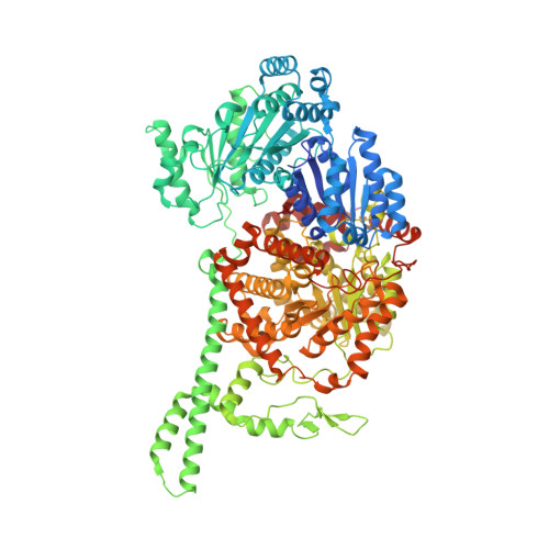
    Acyl-CoA mutases are a growing class of adenosylcobalamin-dependent radical enzymes that perform challenging carbon skeleton rearrangements in primary and secondary metabolism. Members of this class of enzymes must precisely control substrate positioning to prevent oxidative interception of radical intermediates during catalysis. Our understanding of substrate specificity and catalysis in acyl-CoA mutases, however, is incomplete. Here, we present crystal structures of IcmF, a natural fusion protein variant of isobutyryl-CoA mutase, in complex with the adenosylcobalamin cofactor and four different acyl-CoA substrates. These structures demonstrate how the active site is designed to accommodate the aliphatic acyl chains of each substrate. The structures suggest that a conformational change of the 5'-deoxyadenosyl group from C2'-endo to C3'-endo could contribute to initiation of catalysis. Furthermore, detailed bioinformatic analyses guided by our structural findings identify critical determinants of acyl-CoA mutase substrate specificity and predict new acyl-CoA mutase-catalyzed reactions. These results expand our understanding of the substrate specificity and the catalytic scope of acyl-CoA mutases and could benefit engineering efforts for biotechnological applications ranging from production of biofuels and commercial products to hydrocarbon remediation.


  • Organizational Affiliation
    • Departments of Chemistry, Massachusetts Institute of Technology, Cambridge, Massachusetts 02139.

Macromolecule Content 

  • Total Structure Weight: 251.45 kDa 
  • Atom Count: 16,519 
  • Modeled Residue Count: 2,120 
  • Deposited Residue Count: 2,226 
  • Unique protein chains: 1

Macromolecules

Find similar proteins by:|  3D Structure
Entity ID: 1
MoleculeChains  Sequence LengthOrganismDetailsImage
Isobutyryl-CoA mutase fused
A, B
1,113Cupriavidus metallidurans CH34Mutation(s): 0 
Gene Names: IcmFRmet_0210
EC: 5.4.99.2 (PDB Primary Data), 3.6.5 (UniProt), 5.4.99.13 (UniProt)
UniProt
Find proteins for Q1LRY0 (Cupriavidus metallidurans (strain ATCC 43123 / DSM 2839 / NBRC 102507 / CH34))
Explore Q1LRY0 
Go to UniProtKB:  Q1LRY0
Entity Groups
Sequence Clusters30% Identity50% Identity70% Identity90% Identity95% Identity100% Identity
UniProt GroupQ1LRY0
Sequence Annotations
Expand
Reference Sequence

Small Molecules

Ligands 5 Unique
IDChains Name / Formula / InChI Key2D Diagram3D Interactions
B12

Query on B12


Download:Ideal Coordinates CCD File
C [auth A],
I [auth B]
COBALAMIN
C62 H89 Co N13 O14 P
LKVIQTCSMMVGFU-DWSMJLPVSA-N
IVC

Query on IVC


Download:Ideal Coordinates CCD File
E [auth A],
J [auth B]
Isovaleryl-coenzyme A
C26 H44 N7 O17 P3 S
UYVZIWWBJMYRCD-ZMHDXICWSA-N
GDP

Query on GDP


Download:Ideal Coordinates CCD File
F [auth A],
K [auth B]
GUANOSINE-5'-DIPHOSPHATE
C10 H15 N5 O11 P2
QGWNDRXFNXRZMB-UUOKFMHZSA-N
5AD

Query on 5AD


Download:Ideal Coordinates CCD File
D [auth A]5'-DEOXYADENOSINE
C10 H13 N5 O3
XGYIMTFOTBMPFP-KQYNXXCUSA-N
MG

Query on MG


Download:Ideal Coordinates CCD File
G [auth A],
H [auth A],
L [auth B],
M [auth B]
MAGNESIUM ION
Mg
JLVVSXFLKOJNIY-UHFFFAOYSA-N

Experimental Data & Validation

Experimental Data

  • Method: X-RAY DIFFRACTION
  • Resolution: 3.45 Å
  • R-Value Free:  0.214 (Depositor), 0.217 (DCC) 
  • R-Value Work:  0.193 (Depositor), 0.198 (DCC) 
  • R-Value Observed: 0.194 (Depositor) 
Space Group: H 3 2
Unit Cell:
Length ( Å )Angle ( ˚ )
a = 317.56α = 90
b = 317.56β = 90
c = 343.52γ = 120
Software Package:
Software NamePurpose
PHENIXrefinement
XDSdata reduction
XSCALEdata scaling
Cootmodel building

Structure Validation

View Full Validation Report


Ligand Structure Quality Assessment 


Entry History 

& Funding Information

Deposition Data


Funding OrganizationLocationGrant Number
National Institutes of Health/National Institute of General Medical Sciences (NIH/NIGMS)United StatesGM069857
Howard Hughes Medical Institute (HHMI)United States--

Revision History  (Full details and data files)

  • Version 1.0: 2015-09-09
    Type: Initial release
  • Version 1.1: 2015-09-16
    Changes: Database references
  • Version 1.2: 2015-11-18
    Changes: Database references
  • Version 1.3: 2017-09-13
    Changes: Author supporting evidence, Database references, Derived calculations
  • Version 1.4: 2019-11-20
    Changes: Author supporting evidence
  • Version 1.5: 2023-09-27
    Changes: Data collection, Database references, Derived calculations, Refinement description