5CDF

Structure at 2.3 A of the alpha/beta monomer of the F-ATPase from Paracoccus denitrificans


Experimental Data Snapshot

  • Method: X-RAY DIFFRACTION
  • Resolution: 2.30 Å
  • R-Value Free: 0.257 
  • R-Value Work: 0.225 
  • R-Value Observed: 0.227 

wwPDB Validation   3D Report Full Report


This is version 1.2 of the entry. See complete history


Literature

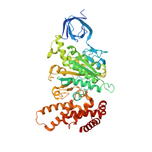
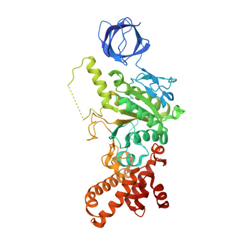
Structure of a catalytic dimer of the alpha- and beta-subunits of the F-ATPase from Paracoccus denitrificans at 2.3 angstrom resolution.

Morales-Rios, E.Montgomery, M.G.Leslie, A.G.Garcia-Trejo, J.J.Walker, J.E.

(2015) Acta Crystallogr F Struct Biol Commun 71: 1309-1317

  • DOI: https://doi.org/10.1107/S2053230X15016076
  • Primary Citation of Related Structures:  
    5CDF

  • PubMed Abstract: 

    The structures of F-ATPases have predominantly been determined from mitochondrial enzymes, and those of the enzymes in eubacteria have been less studied. Paracoccus denitrificans is a member of the α-proteobacteria and is related to the extinct protomitochondrion that became engulfed by the ancestor of eukaryotic cells. The P. denitrificans F-ATPase is an example of a eubacterial F-ATPase that can carry out ATP synthesis only, whereas many others can catalyse both the synthesis and the hydrolysis of ATP. Inhibition of the ATP hydrolytic activity of the P. denitrificans F-ATPase involves the ζ inhibitor protein, an α-helical protein that binds to the catalytic F1 domain of the enzyme. This domain is a complex of three α-subunits and three β-subunits, and one copy of each of the γ-, δ- and ℇ-subunits. Attempts to crystallize the F1-ζ inhibitor complex yielded crystals of a subcomplex of the catalytic domain containing the α- and β-subunits only. Its structure was determined to 2.3 Å resolution and consists of a heterodimer of one α-subunit and one β-subunit. It has no bound nucleotides, and it corresponds to the `open' or `empty' catalytic interface found in other F-ATPases. The main significance of this structure is that it aids in the determination of the structure of the intact membrane-bound F-ATPase, which has been crystallized.


  • Organizational Affiliation

    The Medical Research Council Mitochondrial Biology Unit, Cambridge Biomedical Campus, Hills Road, Cambridge CB2 0XY, England.


Macromolecules
Find similar proteins by:  (by identity cutoff)  |  3D Structure
Entity ID: 1
MoleculeChains Sequence LengthOrganismDetailsImage
ATP synthase subunit alpha511Paracoccus denitrificansMutation(s): 0 
EC: 3.6.3.14
UniProt
Find proteins for A1B8N8 (Paracoccus denitrificans (strain Pd 1222))
Explore A1B8N8 
Go to UniProtKB:  A1B8N8
Entity Groups  
Sequence Clusters30% Identity50% Identity70% Identity90% Identity95% Identity100% Identity
UniProt GroupA1B8N8
Sequence Annotations
Expand
  • Reference Sequence
Find similar proteins by:  (by identity cutoff)  |  3D Structure
Entity ID: 2
MoleculeChains Sequence LengthOrganismDetailsImage
ATP synthase subunit betaB [auth E]474Paracoccus denitrificansMutation(s): 0 
EC: 3.6.3.14
UniProt
Find proteins for A1B8P0 (Paracoccus denitrificans (strain Pd 1222))
Explore A1B8P0 
Go to UniProtKB:  A1B8P0
Entity Groups  
Sequence Clusters30% Identity50% Identity70% Identity90% Identity95% Identity100% Identity
UniProt GroupA1B8P0
Sequence Annotations
Expand
  • Reference Sequence
Experimental Data & Validation

Experimental Data

  • Method: X-RAY DIFFRACTION
  • Resolution: 2.30 Å
  • R-Value Free: 0.257 
  • R-Value Work: 0.225 
  • R-Value Observed: 0.227 
  • Space Group: P 1 21 1
Unit Cell:
Length ( Å )Angle ( ˚ )
a = 72.55α = 90
b = 102.92β = 112.67
c = 89.18γ = 90
Software Package:
Software NamePurpose
REFMACrefinement
Aimlessdata scaling
PDB_EXTRACTdata extraction
PHASERphasing
iMOSFLMdata reduction

Structure Validation

View Full Validation Report


Entry History & Funding Information

Deposition Data


Funding OrganizationLocationGrant Number
United Kingdom--

Revision History  (Full details and data files)

  • Version 1.0: 2015-10-07
    Type: Initial release
  • Version 1.1: 2015-10-28
    Changes: Database references
  • Version 1.2: 2024-01-10
    Changes: Data collection, Database references, Refinement description