Structural and Biochemical Characterization of a Copper-Binding Mutant of the Organomercurial Lyase MerB: Insight into the Key Role of the Active Site Aspartic Acid in Hg-Carbon Bond Cleavage and Metal Binding Specificity.
Wahba, H.M., Lecoq, L., Stevenson, M., Mansour, A., Cappadocia, L., Lafrance-Vanasse, J., Wilkinson, K.J., Sygusch, J., Wilcox, D.E., Omichinski, J.G.(2016) Biochemistry 55: 1070-1081
- PubMed: 26820485 Search on PubMed
- DOI: https://doi.org/10.1021/acs.biochem.5b01298
- Primary Citation Related Structures:
5C0T, 5C0U, 5C17, 5DSF - PubMed Abstract:
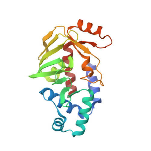
In bacterial resistance to mercury, the organomercurial lyase (MerB) plays a key role in the detoxification pathway through its ability to cleave Hg-carbon bonds. Two cysteines (C96 and C159; Escherichia coli MerB numbering) and an aspartic acid (D99) have been identified as the key catalytic residues, and these three residues are conserved in all but four known MerB variants, where the aspartic acid is replaced with a serine. To understand the role of the active site serine, we characterized the structure and metal binding properties of an E. coli MerB mutant with a serine substituted for D99 (MerB D99S) as well as one of the native MerB variants containing a serine residue in the active site (Bacillus megaterium MerB2). Surprisingly, the MerB D99S protein copurified with a bound metal that was determined to be Cu(II) from UV-vis absorption, inductively coupled plasma mass spectrometry, nuclear magnetic resonance, and electron paramagnetic resonance studies. X-ray structural studies revealed that the Cu(II) is bound to the active site cysteine residues of MerB D99S, but that it is displaced following the addition of either an organomercurial substrate or an ionic mercury product. In contrast, the B. megaterium MerB2 protein does not copurify with copper, but the structure of the B. megaterium MerB2-Hg complex is highly similar to the structure of the MerB D99S-Hg complexes. These results demonstrate that the active site aspartic acid is crucial for both the enzymatic activity and metal binding specificity of MerB proteins and suggest a possible functional relationship between MerB and its only known structural homologue, the copper-binding protein NosL.
- Faculty of Pharmacy, Beni-suef University , Beni-suef, Egypt.
Organizational Affiliation: