5BY8

The structure of Rpf2-Rrs1 explains its role in ribosome biogenesis


Experimental Data Snapshot

  • Method: X-RAY DIFFRACTION
  • Resolution: 1.51 Å
  • R-Value Free: 0.191 
  • R-Value Work: 0.145 
  • R-Value Observed: 0.148 

wwPDB Validation   3D Report Full Report


This is version 1.1 of the entry. See complete history


Literature

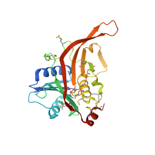
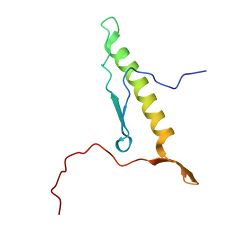
The structure of Rpf2-Rrs1 explains its role in ribosome biogenesis.

Kharde, S.Calvino, F.R.Gumiero, A.Wild, K.Sinning, I.

(2015) Nucleic Acids Res 43: 7083-7095

  • DOI: https://doi.org/10.1093/nar/gkv640
  • Primary Citation of Related Structures:  
    5BY8

  • PubMed Abstract: 

    The assembly of eukaryotic ribosomes is a hierarchical process involving about 200 biogenesis factors and a series of remodeling steps. The 5S RNP consisting of the 5S rRNA, RpL5 and RpL11 is recruited at an early stage, but has to rearrange during maturation of the pre-60S ribosomal subunit. Rpf2 and Rrs1 have been implicated in 5S RNP biogenesis, but their precise role was unclear. Here, we present the crystal structure of the Rpf2-Rrs1 complex from Aspergillus nidulans at 1.5 Å resolution and describe it as Brix domain of Rpf2 completed by Rrs1 to form two anticodon-binding domains with functionally important tails. Fitting the X-ray structure into the cryo-EM density of a previously described pre-60S particle correlates with biochemical data. The heterodimer forms specific contacts with the 5S rRNA, RpL5 and the biogenesis factor Rsa4. The flexible protein tails of Rpf2-Rrs1 localize to the central protuberance. Two helices in the Rrs1 C-terminal tail occupy a strategic position to block the rotation of 25S rRNA and the 5S RNP. Our data provide a structural model for 5S RNP recruitment to the pre-60S particle and explain why removal of Rpf2-Rrs1 is necessary for rearrangements to drive 60S maturation.


  • Organizational Affiliation

    Heidelberg University Biochemistry Center (BZH), INF 328, D-69120 Heidelberg, Germany.


Macromolecules
Find similar proteins by:  (by identity cutoff)  |  3D Structure
Entity ID: 1
MoleculeChains Sequence LengthOrganismDetailsImage
Rpf2231Aspergillus nidulans FGSC A4Mutation(s): 0 
Gene Names: ANIA_10200
UniProt
Find proteins for C8VMF9 (Emericella nidulans (strain FGSC A4 / ATCC 38163 / CBS 112.46 / NRRL 194 / M139))
Explore C8VMF9 
Go to UniProtKB:  C8VMF9
Entity Groups  
Sequence Clusters30% Identity50% Identity70% Identity90% Identity95% Identity100% Identity
UniProt GroupC8VMF9
Sequence Annotations
Expand
  • Reference Sequence
Find similar proteins by:  (by identity cutoff)  |  3D Structure
Entity ID: 2
MoleculeChains Sequence LengthOrganismDetailsImage
Rrs185Aspergillus nidulans FGSC A4Mutation(s): 0 
Gene Names: AN3745.2ANIA_03745
UniProt
Find proteins for Q5B6T5 (Emericella nidulans (strain FGSC A4 / ATCC 38163 / CBS 112.46 / NRRL 194 / M139))
Explore Q5B6T5 
Go to UniProtKB:  Q5B6T5
Entity Groups  
Sequence Clusters30% Identity50% Identity70% Identity90% Identity95% Identity100% Identity
UniProt GroupQ5B6T5
Sequence Annotations
Expand
  • Reference Sequence
Small Molecules
Modified Residues  1 Unique
IDChains TypeFormula2D DiagramParent
MSE
Query on MSE
A
L-PEPTIDE LINKINGC5 H11 N O2 SeMET
Experimental Data & Validation

Experimental Data

  • Method: X-RAY DIFFRACTION
  • Resolution: 1.51 Å
  • R-Value Free: 0.191 
  • R-Value Work: 0.145 
  • R-Value Observed: 0.148 
  • Space Group: C 2 2 21
Unit Cell:
Length ( Å )Angle ( ˚ )
a = 49.16α = 90
b = 84.14β = 90
c = 194.3γ = 90
Software Package:
Software NamePurpose
PHENIXrefinement
XDSdata reduction
XDSdata scaling
PHENIXphasing

Structure Validation

View Full Validation Report


Entry History 

Deposition Data

Revision History  (Full details and data files)

  • Version 1.0: 2015-07-08
    Type: Initial release
  • Version 1.1: 2015-08-26
    Changes: Database references