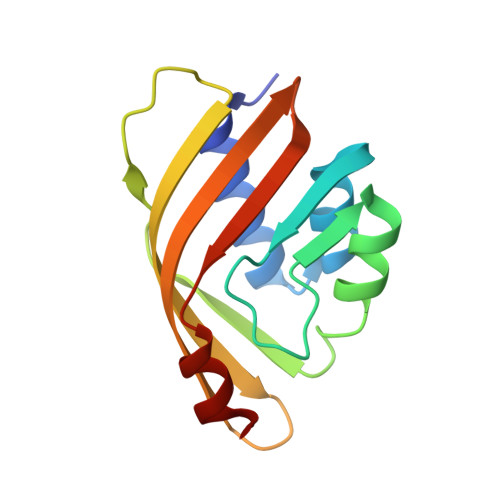
CSAR Benchmark Exercise 2013: Evaluation of Results from a Combined Computational Protein Design, Docking, and Scoring/Ranking Challenge.
Smith, R.D., Damm-Ganamet, K.L., Dunbar, J.B., Ahmed, A., Chinnaswamy, K., Delproposto, J.E., Kubish, G.M., Tinberg, C.E., Khare, S.D., Dou, J., Doyle, L., Stuckey, J.A., Baker, D., Carlson, H.A.(2016) J Chem Inf Model 56: 1022-1031
- PubMed: 26419257
- DOI: https://doi.org/10.1021/acs.jcim.5b00387
- Primary Citation of Related Structures:
5BVB - PubMed Abstract:
Community Structure-Activity Resource (CSAR) conducted a benchmark exercise to evaluate the current computational methods for protein design, ligand docking, and scoring/ranking. The exercise consisted of three phases. The first phase required the participants to identify and rank order which designed sequences were able to bind the small molecule digoxigenin. The second phase challenged the community to select a near-native pose of digoxigenin from a set of decoy poses for two of the designed proteins. The third phase investigated the ability of current methods to rank/score the binding affinity of 10 related steroids to one of the designed proteins (pKd = 4.1 to 6.7). We found that 11 of 13 groups were able to correctly select the sequence that bound digoxigenin, with most groups providing the correct three-dimensional structure for the backbone of the protein as well as all atoms of the active-site residues. Eleven of the 14 groups were able to select the appropriate pose from a set of plausible decoy poses. The ability to predict absolute binding affinities is still a difficult task, as 8 of 14 groups were able to correlate scores to affinity (Pearson-r > 0.7) of the designed protein for congeneric steroids and only 5 of 14 groups were able to correlate the ranks of the 10 related ligands (Spearman-ρ > 0.7).
- Department of Medicinal Chemistry, University of Michigan , 428 Church Street, Ann Arbor, Michigan 48109-1065, United States.
Organizational Affiliation:


