5BO0

Crystal structure of Human MCM2 HBD and ASF1b chaperoning a histone H3.2-H4 dimer


Experimental Data Snapshot

  • Method: X-RAY DIFFRACTION
  • Resolution: 2.91 Å
  • R-Value Free: 0.238 
  • R-Value Work: 0.190 
  • R-Value Observed: 0.193 

wwPDB Validation   3D Report Full Report


This is version 1.4 of the entry. See complete history


Literature

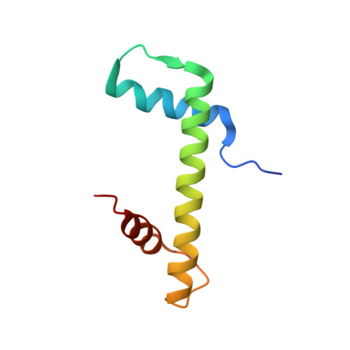
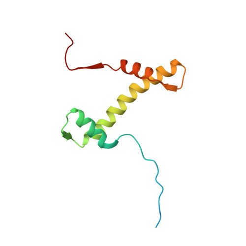
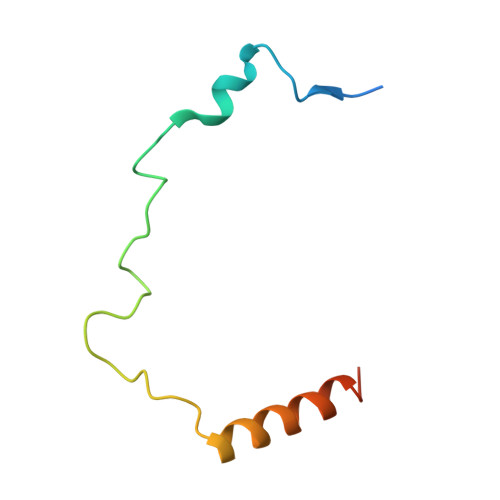
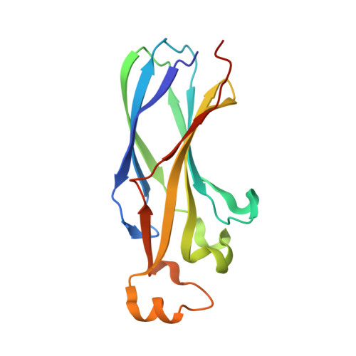
A unique binding mode enables MCM2 to chaperone histones H3-H4 at replication forks.

Huang, H.Strmme, C.B.Saredi, G.Hodl, M.Strandsby, A.Gonzalez-Aguilera, C.Chen, S.Groth, A.Patel, D.J.

(2015) Nat Struct Mol Biol 22: 618-626

  • DOI: https://doi.org/10.1038/nsmb.3055
  • Primary Citation of Related Structures:  
    5BNV, 5BNX, 5BO0

  • PubMed Abstract: 

    During DNA replication, chromatin is reassembled by recycling of modified old histones and deposition of new ones. How histone dynamics integrates with DNA replication to maintain genome and epigenome information remains unclear. Here, we reveal how human MCM2, part of the replicative helicase, chaperones histones H3-H4. Our first structure shows an H3-H4 tetramer bound by two MCM2 histone-binding domains (HBDs), which hijack interaction sites used by nucleosomal DNA. Our second structure reveals MCM2 and ASF1 cochaperoning an H3-H4 dimer. Mutational analyses show that the MCM2 HBD is required for MCM2-7 histone-chaperone function and normal cell proliferation. Further, we show that MCM2 can chaperone both new and old canonical histones H3-H4 as well as H3.3 and CENPA variants. The unique histone-binding mode of MCM2 thus endows the replicative helicase with ideal properties for recycling histones genome wide during DNA replication.


  • Organizational Affiliation

    Structural Biology Program, Memorial Sloan-Kettering Cancer Center, New York, New York, USA.


Macromolecules
Find similar proteins by:  (by identity cutoff)  |  3D Structure
Entity ID: 1
MoleculeChains Sequence LengthOrganismDetailsImage
Histone H3.280Homo sapiensMutation(s): 0 
Gene Names: HIST2H3AHIST2H3CH3F2H3FMHIST2H3D
UniProt & NIH Common Fund Data Resources
Find proteins for Q71DI3 (Homo sapiens)
Explore Q71DI3 
Go to UniProtKB:  Q71DI3
PHAROS:  Q71DI3
Entity Groups  
Sequence Clusters30% Identity50% Identity70% Identity90% Identity95% Identity100% Identity
UniProt GroupQ71DI3
Sequence Annotations
Expand
  • Reference Sequence
Find similar proteins by:  (by identity cutoff)  |  3D Structure
Entity ID: 2
MoleculeChains Sequence LengthOrganismDetailsImage
Histone H4102Homo sapiensMutation(s): 0 
Gene Names: 
UniProt & NIH Common Fund Data Resources
Find proteins for P62805 (Homo sapiens)
Explore P62805 
Go to UniProtKB:  P62805
PHAROS:  P62805
Entity Groups  
Sequence Clusters30% Identity50% Identity70% Identity90% Identity95% Identity100% Identity
UniProt GroupP62805
Sequence Annotations
Expand
  • Reference Sequence
Find similar proteins by:  (by identity cutoff)  |  3D Structure
Entity ID: 3
MoleculeChains Sequence LengthOrganismDetailsImage
DNA replication licensing factor MCM270Homo sapiensMutation(s): 0 
Gene Names: MCM2BM28CCNL1CDCL1KIAA0030
UniProt & NIH Common Fund Data Resources
Find proteins for P49736 (Homo sapiens)
Explore P49736 
Go to UniProtKB:  P49736
PHAROS:  P49736
GTEx:  ENSG00000073111 
Entity Groups  
Sequence Clusters30% Identity50% Identity70% Identity90% Identity95% Identity100% Identity
UniProt GroupP49736
Sequence Annotations
Expand
  • Reference Sequence
Find similar proteins by:  (by identity cutoff)  |  3D Structure
Entity ID: 4
MoleculeChains Sequence LengthOrganismDetailsImage
Histone chaperone ASF1B158Homo sapiensMutation(s): 0 
Gene Names: ASF1B
UniProt & NIH Common Fund Data Resources
Find proteins for Q9NVP2 (Homo sapiens)
Explore Q9NVP2 
Go to UniProtKB:  Q9NVP2
PHAROS:  Q9NVP2
GTEx:  ENSG00000105011 
Entity Groups  
Sequence Clusters30% Identity50% Identity70% Identity90% Identity95% Identity100% Identity
UniProt GroupQ9NVP2
Sequence Annotations
Expand
  • Reference Sequence
Small Molecules
Ligands 1 Unique
IDChains Name / Formula / InChI Key2D Diagram3D Interactions
GOL
Query on GOL

Download Ideal Coordinates CCD File 
E [auth B]GLYCEROL
C3 H8 O3
PEDCQBHIVMGVHV-UHFFFAOYSA-N
Experimental Data & Validation

Experimental Data

  • Method: X-RAY DIFFRACTION
  • Resolution: 2.91 Å
  • R-Value Free: 0.238 
  • R-Value Work: 0.190 
  • R-Value Observed: 0.193 
  • Space Group: P 31 2 1
Unit Cell:
Length ( Å )Angle ( ˚ )
a = 112.964α = 90
b = 112.964β = 90
c = 98.226γ = 120
Software Package:
Software NamePurpose
PHENIXrefinement
HKL-2000data reduction
HKL-2000data scaling
PHASERphasing
Cootmodel building

Structure Validation

View Full Validation Report


Entry History 

Deposition Data

Revision History  (Full details and data files)

  • Version 1.0: 2015-06-17
    Type: Initial release
  • Version 1.1: 2015-07-15
    Changes: Database references
  • Version 1.2: 2015-07-29
    Changes: Database references
  • Version 1.3: 2015-08-19
    Changes: Database references
  • Version 1.4: 2024-03-06
    Changes: Data collection, Database references, Derived calculations