5AYD | pdb_00005ayd

Crystal structure of Ruminococcus albus beta-(1,4)-mannooligosaccharide phosphorylase (RaMP2) in complexes with phosphate


Experimental Data Snapshot

  • Method: X-RAY DIFFRACTION
  • Resolution: 2.30 Å
  • R-Value Free: 
    0.212 (Depositor), 0.213 (DCC) 
  • R-Value Work: 
    0.168 (Depositor), 0.171 (DCC) 
  • R-Value Observed: 
    0.170 (Depositor) 

wwPDB Validation 3D Report Full Report

Validation slider image for 5AYD

This is version 1.3 of the entry. See complete history

Literature

Structural insights into the difference in substrate recognition of two mannoside phosphorylases from two GH130 subfamilies.

Ye, Y.Saburi, W.Odaka, R.Kato, K.Sakurai, N.Komoda, K.Nishimoto, M.Kitaoka, M.Mori, H.Yao, M.

(2016) FEBS Lett 590: 828-837

  • DOI: https://doi.org/10.1002/1873-3468.12105
  • Primary Citation Related Structures: 
    5AY9, 5AYC, 5AYD, 5AYE

  • PubMed Abstract: 

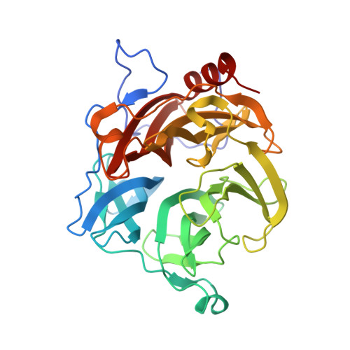
    In Ruminococcus albus, 4-O-β-D-mannosyl-D-glucose phosphorylase (RaMP1) and β-(1,4)-mannooligosaccharide phosphorylase (RaMP2) belong to two subfamilies of glycoside hydrolase family 130. The two enzymes phosphorolyze β-mannosidic linkages at the nonreducing ends of their substrates, and have substantially diverse substrate specificity. The differences in their mechanism of substrate binding have not yet been fully clarified. In the present study, we report the crystal structures of RaMP1 with/without 4-O-β-D-mannosyl-d-glucose and RaMP2 with/without β-(1→4)-mannobiose. The structures of the two enzymes differ at the +1 subsite of the substrate-binding pocket. Three loops are proposed to determine the different substrate specificities. One of these loops is contributed from the adjacent molecule of the oligomer structure. In RaMP1, His245 of loop 3 forms a hydrogen-bond network with the substrate through a water molecule, and is indispensible for substrate binding.


  • Organizational Affiliation
    • Graduate School of Life Science, Hokkaido University, Sapporo, Japan.

Macromolecule Content 

  • Total Structure Weight: 230.68 kDa 
  • Atom Count: 17,442 
  • Modeled Residue Count: 2,010 
  • Deposited Residue Count: 2,010 
  • Unique protein chains: 1

Macromolecules

Find similar proteins by:|  3D Structure
Entity ID: 1
MoleculeChains  Sequence LengthOrganismDetailsImage
Beta-1,4-mannooligosaccharide phosphorylase
A, B, C, D, E
A, B, C, D, E, F
335Hominimerdicola alba 7 = DSM 20455Mutation(s): 0 
Gene Names: Rumal_0099
EC: 2.4.1.319
UniProt
Find proteins for E6UBR9 (Ruminococcus albus (strain ATCC 27210 / DSM 20455 / JCM 14654 / NCDO 2250 / 7))
Explore E6UBR9 
Go to UniProtKB:  E6UBR9
Entity Groups
Sequence Clusters30% Identity50% Identity70% Identity90% Identity95% Identity100% Identity
UniProt GroupE6UBR9
Sequence Annotations
Expand
Reference Sequence

Experimental Data & Validation

Experimental Data

  • Method: X-RAY DIFFRACTION
  • Resolution: 2.30 Å
  • R-Value Free:  0.212 (Depositor), 0.213 (DCC) 
  • R-Value Work:  0.168 (Depositor), 0.171 (DCC) 
  • R-Value Observed: 0.170 (Depositor) 
Space Group: P 1 21 1
Unit Cell:
Length ( Å )Angle ( ˚ )
a = 92.678α = 90
b = 166.181β = 118.91
c = 92.969γ = 90
Software Package:
Software NamePurpose
PHENIXrefinement
XDSdata processing
Cootrefinement
PHENIXmodel building

Structure Validation

View Full Validation Report


Entry History 

Deposition Data

Revision History  (Full details and data files)

  • Version 1.0: 2016-03-23
    Type: Initial release
  • Version 1.1: 2016-04-06
    Changes: Database references
  • Version 1.2: 2020-02-26
    Changes: Data collection, Database references, Derived calculations
  • Version 1.3: 2024-03-20
    Changes: Data collection, Database references