5AKB

MutS in complex with the N-terminal domain of MutL - crystal form 1


Experimental Data Snapshot

  • Method: X-RAY DIFFRACTION
  • Resolution: 4.71 Å
  • R-Value Free: 0.349 
  • R-Value Work: 0.319 
  • R-Value Observed: 0.321 

wwPDB Validation   3D Report Full Report


Ligand Structure Quality Assessment 


This is version 1.2 of the entry. See complete history


Literature

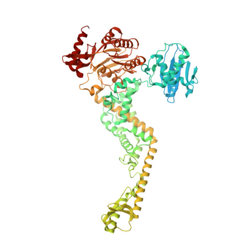
MutS/MutL crystal structure reveals that the MutS sliding clamp loads MutL onto DNA.

Groothuizen, F.S.Winkler, I.Cristovao, M.Fish, A.Winterwerp, H.H.Reumer, A.Marx, A.D.Hermans, N.Nicholls, R.A.Murshudov, G.N.Lebbink, J.H.Friedhoff, P.Sixma, T.K.

(2015) Elife 4: e06744-e06744

  • DOI: https://doi.org/10.7554/eLife.06744
  • Primary Citation of Related Structures:  
    5AKB, 5AKC, 5AKD

  • PubMed Abstract: 

    To avoid mutations in the genome, DNA replication is generally followed by DNA mismatch repair (MMR). MMR starts when a MutS homolog recognizes a mismatch and undergoes an ATP-dependent transformation to an elusive sliding clamp state. How this transient state promotes MutL homolog recruitment and activation of repair is unclear. Here we present a crystal structure of the MutS/MutL complex using a site-specifically crosslinked complex and examine how large conformational changes lead to activation of MutL. The structure captures MutS in the sliding clamp conformation, where tilting of the MutS subunits across each other pushes DNA into a new channel, and reorientation of the connector domain creates an interface for MutL with both MutS subunits. Our work explains how the sliding clamp promotes loading of MutL onto DNA, to activate downstream effectors. We thus elucidate a crucial mechanism that ensures that MMR is initiated only after detection of a DNA mismatch.


  • Organizational Affiliation

    Division of Biochemistry and CGC.nl, Netherlands Cancer Institute, Amsterdam, Netherlands.


Macromolecules
Find similar proteins by:  (by identity cutoff)  |  3D Structure
Entity ID: 1
MoleculeChains Sequence LengthOrganismDetailsImage
DNA MISMATCH REPAIR PROTEIN MUTS
A, B, E
800Escherichia coli K-12Mutation(s): 7 
UniProt
Find proteins for P23909 (Escherichia coli (strain K12))
Explore P23909 
Go to UniProtKB:  P23909
Entity Groups  
Sequence Clusters30% Identity50% Identity70% Identity90% Identity95% Identity100% Identity
UniProt GroupP23909
Sequence Annotations
Expand
  • Reference Sequence
Find similar proteins by:  (by identity cutoff)  |  3D Structure
Entity ID: 2
MoleculeChains Sequence LengthOrganismDetailsImage
DNA MISMATCH REPAIR PROTEIN MUTL
C, D, F
369Escherichia coli K-12Mutation(s): 5 
UniProt
Find proteins for P23367 (Escherichia coli (strain K12))
Explore P23367 
Go to UniProtKB:  P23367
Entity Groups  
Sequence Clusters30% Identity50% Identity70% Identity90% Identity95% Identity100% Identity
UniProt GroupP23367
Sequence Annotations
Expand
  • Reference Sequence
Experimental Data & Validation

Experimental Data

  • Method: X-RAY DIFFRACTION
  • Resolution: 4.71 Å
  • R-Value Free: 0.349 
  • R-Value Work: 0.319 
  • R-Value Observed: 0.321 
  • Space Group: C 1 2 1
Unit Cell:
Length ( Å )Angle ( ˚ )
a = 165.932α = 90
b = 188.544β = 94.77
c = 200.426γ = 90
Software Package:
Software NamePurpose
REFMACrefinement
XDSdata reduction
Aimlessdata scaling
PHASERphasing

Structure Validation

View Full Validation Report


Ligand Structure Quality Assessment 


Entry History 

Revision History  (Full details and data files)

  • Version 1.0: 2015-07-22
    Type: Initial release
  • Version 1.1: 2019-02-27
    Changes: Data collection, Database references, Other
  • Version 1.2: 2024-01-10
    Changes: Data collection, Database references, Derived calculations, Other, Refinement description