5A6Z

Structure of the LecB lectin from Pseudomonas aeruginosa strain PA14 in complex with lewis a


Experimental Data Snapshot

  • Method: X-RAY DIFFRACTION
  • Resolution: 1.50 Å
  • R-Value Free: 0.155 
  • R-Value Work: 0.131 
  • R-Value Observed: 0.132 

wwPDB Validation   3D Report Full Report


This is version 2.1 of the entry. See complete history


Literature

The virulence factor LecB varies in clinical isolates: consequences for ligand binding and drug discovery.

Sommer, R.Wagner, S.Varrot, A.Nycholat, C.M.Khaledi, A.Haussler, S.Paulson, J.C.Imberty, A.Titz, A.

(2016) Chem Sci 7: 4990-5001

  • DOI: https://doi.org/10.1039/c6sc00696e
  • Primary Citation of Related Structures:  
    5A6Q, 5A6X, 5A6Y, 5A6Z

  • PubMed Abstract: 

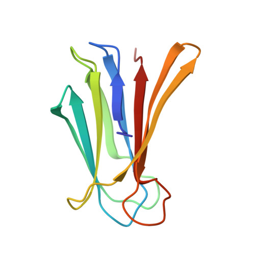
    P. aeruginosa causes a substantial number of nosocomial infections and is the leading cause of death of cystic fibrosis patients. This Gram-negative bacterium is highly resistant against antibiotics and further protects itself by forming a biofilm. Moreover, a high genomic variability among clinical isolates complicates therapy. Its lectin LecB is a virulence factor and necessary for adhesion and biofilm formation. We analyzed the sequence of LecB variants in a library of clinical isolates and demonstrate that it can serve as a marker for strain family classification. LecB from the highly virulent model strain PA14 presents 13% sequence divergence with LecB from the well characterized PAO1 strain. These differences might result in differing ligand binding specificities and ultimately in reduced efficacy of drugs directed towards LecB. Despite several amino acid variations at the carbohydrate binding site, glycan array analysis showed a comparable binding pattern for both variants. A common high affinity ligand could be identified and after its chemoenzymatic synthesis verified in a competitive binding assay: an N -glycan presenting two blood group O epitopes (H-type 2 antigen). Molecular modeling of the complex suggests a bivalent interaction of the ligand with the LecB tetramer by bridging two separate binding sites. This binding rationalizes the strong avidity (35 nM) of LecB PA14 to this human fucosylated N-glycan. Biochemical evaluation of a panel of glycan ligands revealed that LecB PA14 demonstrated higher glycan affinity compared to LecB PAO1 including the extraordinarily potent affinity of 70 nM towards the monovalent human antigen Lewis a . The structural basis of this unusual high affinity ligand binding for lectins was rationalized by solving the protein crystal structures of LecB PA14 with several glycans.


  • Organizational Affiliation

    Chemical Biology of Carbohydrates , Helmholtz Institute for Pharmaceutical Research Saarland (HIPS) , D-66123 Saarbrücken , Germany . Email: alexander.titz@helmholtz-hzi.de ; http://www.helmholtz-hzi.de/cbch.


Macromolecules
Find similar proteins by:  (by identity cutoff)  |  3D Structure
Entity ID: 1
MoleculeChains Sequence LengthOrganismDetailsImage
LECB
A, B, C, D
114Pseudomonas aeruginosa UCBPP-PA14Mutation(s): 0 
UniProt
Find proteins for A0A0H2ZE85 (Pseudomonas aeruginosa (strain UCBPP-PA14))
Explore A0A0H2ZE85 
Go to UniProtKB:  A0A0H2ZE85
Entity Groups  
Sequence Clusters30% Identity50% Identity70% Identity90% Identity95% Identity100% Identity
UniProt GroupA0A0H2ZE85
Sequence Annotations
Expand
  • Reference Sequence
Oligosaccharides

Help

Entity ID: 2
MoleculeChains Length2D Diagram Glycosylation3D Interactions
beta-D-galactopyranose-(1-3)-[alpha-L-fucopyranose-(1-4)]2-acetamido-2-deoxy-beta-D-glucopyranose-(1-3)-alpha-D-galactopyranose
E, H
4N/A
Glycosylation Resources
GlyTouCan:  G13393XR
GlyCosmos:  G13393XR
GlyGen:  G13393XR
Entity ID: 3
MoleculeChains Length2D Diagram Glycosylation3D Interactions
beta-D-galactopyranose-(1-3)-[alpha-L-fucopyranose-(1-4)]2-acetamido-2-deoxy-beta-D-glucopyranose
F
3N/A
Glycosylation Resources
GlyTouCan:  G39023AU
GlyCosmos:  G39023AU
GlyGen:  G39023AU
Entity ID: 4
MoleculeChains Length2D Diagram Glycosylation3D Interactions
beta-D-galactopyranose-(1-3)-[alpha-L-fucopyranose-(1-4)]2-acetamido-2-deoxy-beta-D-glucopyranose-(1-3)-beta-D-galactopyranose
G
4N/A
Glycosylation Resources
GlyTouCan:  G80022XC
GlyCosmos:  G80022XC
GlyGen:  G80022XC
Small Molecules
Ligands 3 Unique
IDChains Name / Formula / InChI Key2D Diagram3D Interactions
PEG
Query on PEG

Download Ideal Coordinates CCD File 
T [auth D]DI(HYDROXYETHYL)ETHER
C4 H10 O3
MTHSVFCYNBDYFN-UHFFFAOYSA-N
SO4
Query on SO4

Download Ideal Coordinates CCD File 
K [auth A],
N [auth B],
Q [auth C]
SULFATE ION
O4 S
QAOWNCQODCNURD-UHFFFAOYSA-L
CA
Query on CA

Download Ideal Coordinates CCD File 
I [auth A]
J [auth A]
L [auth B]
M [auth B]
O [auth C]
I [auth A],
J [auth A],
L [auth B],
M [auth B],
O [auth C],
P [auth C],
R [auth D],
S [auth D]
CALCIUM ION
Ca
BHPQYMZQTOCNFJ-UHFFFAOYSA-N
Experimental Data & Validation

Experimental Data

  • Method: X-RAY DIFFRACTION
  • Resolution: 1.50 Å
  • R-Value Free: 0.155 
  • R-Value Work: 0.131 
  • R-Value Observed: 0.132 
  • Space Group: P 1 21 1
Unit Cell:
Length ( Å )Angle ( ˚ )
a = 52.976α = 90
b = 63.772β = 91.25
c = 64.513γ = 90
Software Package:
Software NamePurpose
REFMACrefinement
XDSdata reduction
Aimlessdata scaling
PHASERphasing

Structure Validation

View Full Validation Report


Entry History 

Deposition Data

Revision History  (Full details and data files)

  • Version 1.0: 2016-05-25
    Type: Initial release
  • Version 1.1: 2018-09-12
    Changes: Data collection, Database references
  • Version 1.2: 2020-01-22
    Changes: Data collection, Derived calculations, Other, Structure summary
  • Version 2.0: 2020-07-29
    Type: Remediation
    Reason: Carbohydrate remediation
    Changes: Advisory, Atomic model, Data collection, Derived calculations, Structure summary
  • Version 2.1: 2024-01-10
    Changes: Data collection, Database references, Derived calculations, Refinement description, Structure summary