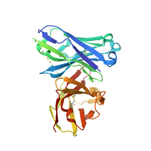
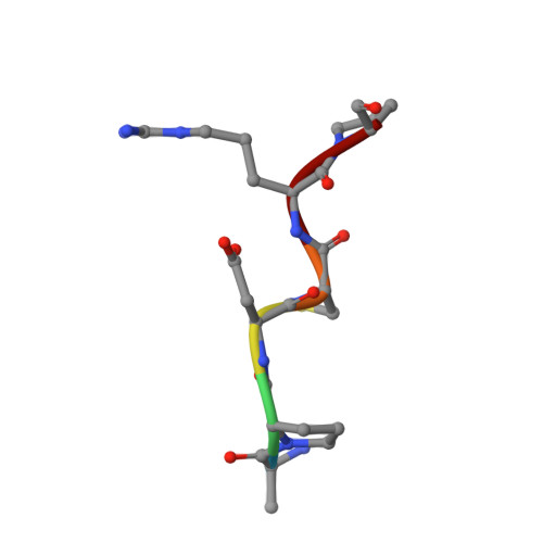
5A2L

Crystal structure of scFv-SM3 in complex with APD-CGalNAc-RP


Experimental Data Snapshot

  • Method: X-RAY DIFFRACTION
  • Resolution: 1.79 Å
  • R-Value Free: 0.204 
  • R-Value Work: 0.184 
  • R-Value Observed: 0.185 

Starting Model: experimental
View more details

wwPDB Validation   3D Report Full Report


Ligand Structure Quality Assessment 


This is version 2.1 of the entry. See complete history


Literature

Deciphering the Non-Equivalence of Serine and Threonine O-Glycosylation Points: Implications for Molecular Recognition of the Tn Antigen by an Anti-Muc1 Antibody.

Martinez-Saez, N.Castro-Lopez, J.Valero-Gonzalez, J.Madariaga, D.Companon, I.Somovilla, V.J.Salvado, M.Asensio, J.L.Jimenez-Barbero, J.Avenoza, A.Busto, J.H.Bernardes, G.J.L.Peregrina, J.M.Hurtado-Guerrero, R.Corzana, F.

(2015) Angew Chem Int Ed Engl 54: 9830

  • DOI: https://doi.org/10.1002/anie.201502813
  • Primary Citation of Related Structures:  
    5A2I, 5A2J, 5A2K, 5A2L

  • PubMed Abstract: 

    The structural features of MUC1-like glycopeptides bearing the Tn antigen (α-O-GalNAc-Ser/Thr) in complex with an anti MUC-1 antibody are reported at atomic resolution. For the α-O-GalNAc-Ser derivative, the glycosidic linkage adopts a high-energy conformation, barely populated in the free state. This unusual structure (also observed in an α-S-GalNAc-Cys mimic) is stabilized by hydrogen bonds between the peptidic fragment and the sugar. The selection of a particular peptide structure by the antibody is thus propagated to the carbohydrate through carbohydrate/peptide contacts, which force a change in the orientation of the sugar moiety. This seems to be unfeasible in the α-O-GalNAc-Thr glycopeptide owing to the more limited flexibility of the side chain imposed by the methyl group. Our data demonstrate the non-equivalence of Ser and Thr O-glycosylation points in molecular recognition processes. These features provide insight into the occurrence in nature of the APDTRP epitope for anti-MUC1 antibodies.


  • Organizational Affiliation

    Departamento de Química, Universidad de La Rioja, Centro de Investigación en Síntesis Química, 26006 Logroño (Spain).


Macromolecules
Find similar proteins by:  (by identity cutoff)  |  3D Structure
Entity ID: 1
MoleculeChains Sequence LengthOrganismDetailsImage
SCFV-SM3A [auth H]244Mus musculusMutation(s): 0 
UniProt
Find proteins for P01727 (Mus musculus)
Explore P01727 
Go to UniProtKB:  P01727
Find proteins for P01801 (Mus musculus)
Explore P01801 
Go to UniProtKB:  P01801
Entity Groups  
Sequence Clusters30% Identity50% Identity70% Identity90% Identity95% Identity100% Identity
UniProt GroupsP01801P01727
Sequence Annotations
Expand
  • Reference Sequence

Find similar proteins by:  Sequence   |   3D Structure  

Entity ID: 2
MoleculeChains Sequence LengthOrganismDetailsImage
MODIFIED ANTIGEN TNB [auth P]6Homo sapiensMutation(s): 0 
Glycosylation
Glycosylation Sites: 1
Sequence Annotations
Expand
  • Reference Sequence
Experimental Data & Validation

Experimental Data

  • Method: X-RAY DIFFRACTION
  • Resolution: 1.79 Å
  • R-Value Free: 0.204 
  • R-Value Work: 0.184 
  • R-Value Observed: 0.185 
  • Space Group: P 1 21 1
Unit Cell:
Length ( Å )Angle ( ˚ )
a = 46.08α = 90
b = 35.19β = 103.01
c = 66.29γ = 90
Software Package:
Software NamePurpose
REFMACrefinement
XDSdata reduction
SCALEPACKdata scaling
MOLREPphasing

Structure Validation

View Full Validation Report


Ligand Structure Quality Assessment 


Entry History 

Revision History  (Full details and data files)

  • Version 1.0: 2015-06-03
    Type: Initial release
  • Version 1.1: 2015-07-08
    Changes: Database references
  • Version 1.2: 2015-08-26
    Changes: Database references
  • Version 2.0: 2020-07-29
    Type: Remediation
    Reason: Carbohydrate remediation
    Changes: Atomic model, Data collection, Derived calculations, Other, Structure summary
  • Version 2.1: 2024-01-10
    Changes: Data collection, Database references, Refinement description, Structure summary