


The Structure of MT189-Tubulin Complex Provides Insights into Drug Design
Li, Z.P., Ma, L.L., Wu, C.Y., Meng, T., Ma, L.P., Zheng, W., Yu, Y., Chen, Q., Yang, J.L., Shen, J.G.(2019) Lett Drug Des Discov
Experimental Data Snapshot
(2019) Lett Drug Des Discov
Entity ID: 1 | |||||
|---|---|---|---|---|---|
| Molecule | Chains | Sequence Length | Organism | Details | Image |
| Tubulin alpha-1B chain | 450 | Sus scrofa | Mutation(s): 0 Gene Names: TUBA1B EC: 3.6.5 |  | |
UniProt | |||||
Entity Groups | |||||
| Sequence Clusters | 30% Identity50% Identity70% Identity90% Identity95% Identity100% Identity | ||||
| UniProt Group | Q2XVP4 | ||||
Sequence AnnotationsExpand | |||||
Reference Sequence | |||||
Entity ID: 2 | |||||
|---|---|---|---|---|---|
| Molecule | Chains | Sequence Length | Organism | Details | Image |
| Tubulin beta-2B chain | 445 | Bos taurus | Mutation(s): 2 Gene Names: TUBB2B | 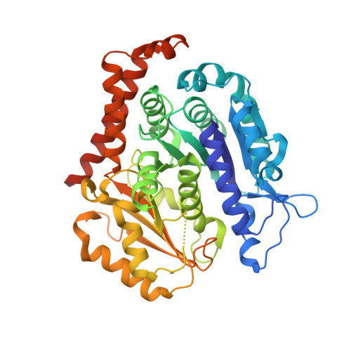 | |
UniProt | |||||
Entity Groups | |||||
| Sequence Clusters | 30% Identity50% Identity70% Identity90% Identity95% Identity100% Identity | ||||
| UniProt Group | Q6B856 | ||||
Sequence AnnotationsExpand | |||||
Reference Sequence | |||||
Entity ID: 3 | |||||
|---|---|---|---|---|---|
| Molecule | Chains | Sequence Length | Organism | Details | Image |
| Stathmin-4 | 143 | Rattus norvegicus | Mutation(s): 0 Gene Names: Stmn4 |  | |
UniProt | |||||
Entity Groups | |||||
| Sequence Clusters | 30% Identity50% Identity70% Identity90% Identity95% Identity100% Identity | ||||
| UniProt Group | P63043 | ||||
Sequence AnnotationsExpand | |||||
Reference Sequence | |||||
Entity ID: 4 | |||||
|---|---|---|---|---|---|
| Molecule | Chains | Sequence Length | Organism | Details | Image |
| Tubulin tyrosine ligase | 384 | Gallus gallus | Mutation(s): 0 Gene Names: TTL | 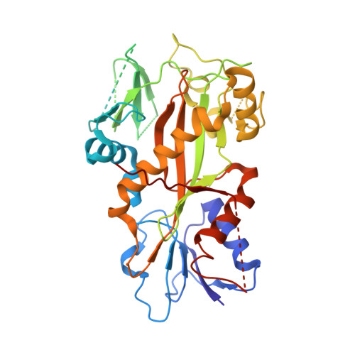 | |
| Ligands 7 Unique | |||||
|---|---|---|---|---|---|
| ID | Chains | Name / Formula / InChI Key | 2D Diagram | 3D Interactions | |
| GTP Download:Ideal Coordinates CCD File | G [auth A], P [auth C], S [auth D] | GUANOSINE-5'-TRIPHOSPHATE C10 H16 N5 O14 P3 XKMLYUALXHKNFT-UUOKFMHZSA-N |  | ||
| ACP Download:Ideal Coordinates CCD File | V [auth F] | PHOSPHOMETHYLPHOSPHONIC ACID ADENYLATE ESTER C11 H18 N5 O12 P3 UFZTZBNSLXELAL-IOSLPCCCSA-N |  | ||
| GDP Download:Ideal Coordinates CCD File | J [auth B] | GUANOSINE-5'-DIPHOSPHATE C10 H15 N5 O11 P2 QGWNDRXFNXRZMB-UUOKFMHZSA-N |  | ||
| 9LX (Subject of Investigation/LOI) Download:Ideal Coordinates CCD File | O [auth B], U [auth D] | 2-(6-fluoro-3-{[(4-methoxyphenyl)methyl]amino}imidazo[1,2-a]pyridin-2-yl)phenol C21 H18 F N3 O2 QBNMSKFVSPKNSV-UHFFFAOYSA-N |  | ||
| MES Download:Ideal Coordinates CCD File | L [auth B], N [auth B] | 2-(N-MORPHOLINO)-ETHANESULFONIC ACID C6 H13 N O4 S SXGZJKUKBWWHRA-UHFFFAOYSA-N |  | ||
| CA Download:Ideal Coordinates CCD File | I [auth A], M [auth B], R [auth C] | CALCIUM ION Ca BHPQYMZQTOCNFJ-UHFFFAOYSA-N |  | ||
| MG Download:Ideal Coordinates CCD File | H [auth A], K [auth B], Q [auth C], T [auth D] | MAGNESIUM ION Mg JLVVSXFLKOJNIY-UHFFFAOYSA-N |  | ||
| Length ( Å ) | Angle ( ˚ ) |
|---|---|
| a = 104.91 | α = 90 |
| b = 157.06 | β = 90 |
| c = 182.838 | γ = 90 |
| Software Name | Purpose |
|---|---|
| REFMAC | refinement |
| HKL-2000 | data reduction |
| HKL-2000 | data scaling |
| PHASER | phasing |


