5ZQY

Crystal structure of a poly(ADP-ribose) glycohydrolase


Experimental Data Snapshot

  • Method: X-RAY DIFFRACTION
  • Resolution: 1.58 Å
  • R-Value Free: 0.180 
  • R-Value Work: 0.157 
  • R-Value Observed: 0.159 

wwPDB Validation   3D Report Full Report


Ligand Structure Quality Assessment 


This is version 1.2 of the entry. See complete history


Literature

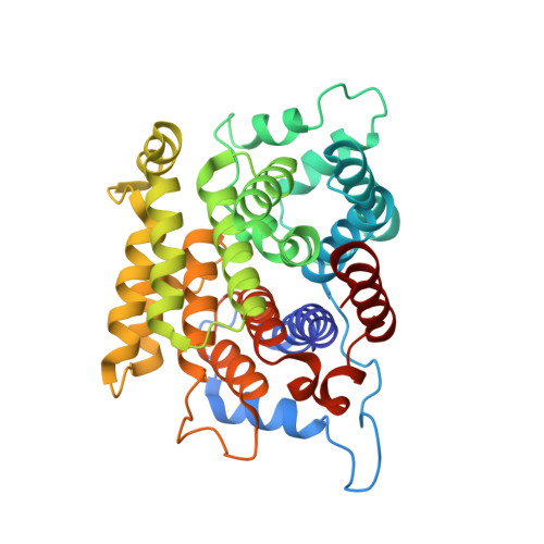
Structure-function analyses reveal the mechanism of the ARH3-dependent hydrolysis of ADP-ribosylation.

Wang, M.Yuan, Z.Xie, R.Ma, Y.Liu, X.Yu, X.

(2018) J Biol Chem 293: 14470-14480

  • DOI: https://doi.org/10.1074/jbc.RA118.004284
  • Primary Citation of Related Structures:  
    5ZQY

  • PubMed Abstract: 

    ADP-ribosylation of proteins plays key roles in multiple biological processes, including DNA damage repair. Recent evidence suggests that serine is an important acceptor for ADP-ribosylation, and that serine ADP-ribosylation is hydrolyzed by ADP-ribosylhydrolase 3 (ARH3 or ADPRHL2). However, the structural details in ARH3-mediated hydrolysis remain elusive. Here, we determined the structure of ARH3 in a complex with ADP-ribose (ADPR). Our analyses revealed a group of acidic residues in ARH3 that keep two Mg 2+ ions at the catalytic center for hydrolysis of Ser-linked ADP-ribosyl group. In particular, dynamic conformational changes involving Glu 41 were observed in the catalytic center. Our observations suggest that Mg 2+ ions together with Glu 41 and water351 are likely to mediate the cleavage of the glycosidic bond in the serine-ADPR substrate. Moreover, we found that ADPR is buried in a groove and forms multiple hydrogen bonds with the main chain and side chains of ARH3 residues. On the basis of these structural findings, we used site-directed mutagenesis to examine the functional roles of key residues in the catalytic pocket of ARH3 in mediating the hydrolysis of ADP-ribosyl from serine and DNA damage repair. Moreover, we noted that ADPR recognition is essential for the recruitment of ARH3 to DNA lesions. Taken together, our study provides structural and functional insights into the molecular mechanism by which ARH3 hydrolyzes the ADP-ribosyl group from serine and contributes to DNA damage repair.


  • Organizational Affiliation

    From the College of Life Sciences, Hebei University, Baoding, 071000 Hebei, China.


Macromolecules
Find similar proteins by:  (by identity cutoff)  |  3D Structure
Entity ID: 1
MoleculeChains Sequence LengthOrganismDetailsImage
Poly(ADP-ribose) glycohydrolase ARH3349Homo sapiensMutation(s): 0 
Gene Names: ADPRHL2ARH3
EC: 3.2.1.143
UniProt & NIH Common Fund Data Resources
Find proteins for Q9NX46 (Homo sapiens)
Explore Q9NX46 
Go to UniProtKB:  Q9NX46
PHAROS:  Q9NX46
GTEx:  ENSG00000116863 
Entity Groups  
Sequence Clusters30% Identity50% Identity70% Identity90% Identity95% Identity100% Identity
UniProt GroupQ9NX46
Sequence Annotations
Expand
  • Reference Sequence
Small Molecules
Ligands 2 Unique
IDChains Name / Formula / InChI Key2D Diagram3D Interactions
AR6
Query on AR6

Download Ideal Coordinates CCD File 
D [auth A][(2R,3S,4R,5R)-5-(6-AMINOPURIN-9-YL)-3,4-DIHYDROXY-OXOLAN-2-YL]METHYL [HYDROXY-[[(2R,3S,4R,5S)-3,4,5-TRIHYDROXYOXOLAN-2-YL]METHOXY]PHOSPHORYL] HYDROGEN PHOSPHATE
C15 H23 N5 O14 P2
SRNWOUGRCWSEMX-ZQSHOCFMSA-N
MG
Query on MG

Download Ideal Coordinates CCD File 
B [auth A],
C [auth A]
MAGNESIUM ION
Mg
JLVVSXFLKOJNIY-UHFFFAOYSA-N
Experimental Data & Validation

Experimental Data

  • Method: X-RAY DIFFRACTION
  • Resolution: 1.58 Å
  • R-Value Free: 0.180 
  • R-Value Work: 0.157 
  • R-Value Observed: 0.159 
  • Space Group: P 1 21 1
Unit Cell:
Length ( Å )Angle ( ˚ )
a = 43.88α = 90
b = 76.74β = 103
c = 48.53γ = 90
Software Package:
Software NamePurpose
PHENIXrefinement
HKL-2000data reduction
HKL-2000data scaling
PHENIXphasing

Structure Validation

View Full Validation Report


Ligand Structure Quality Assessment 


Entry History & Funding Information

Deposition Data


Funding OrganizationLocationGrant Number
National Natural Science Foundation of ChinaChina81672794

Revision History  (Full details and data files)

  • Version 1.0: 2018-08-15
    Type: Initial release
  • Version 1.1: 2018-09-26
    Changes: Data collection, Database references
  • Version 1.2: 2023-11-22
    Changes: Data collection, Database references, Derived calculations, Refinement description, Structure summary