5ZMJ | pdb_00005zmj

Crystal structure of the Fab region of a neutralizing fully human antibody against GM-CSF


Experimental Data Snapshot

  • Method: X-RAY DIFFRACTION
  • Resolution: 1.81 Å
  • R-Value Free: 
    0.191 (Depositor), 0.213 (DCC) 
  • R-Value Work: 
    0.161 (Depositor) 
  • R-Value Observed: 
    0.163 (Depositor) 

Starting Model: experimental
View more details

wwPDB Validation 3D Report Full Report

Validation slider image for 5ZMJ

This is version 1.3 of the entry. See complete history

Literature

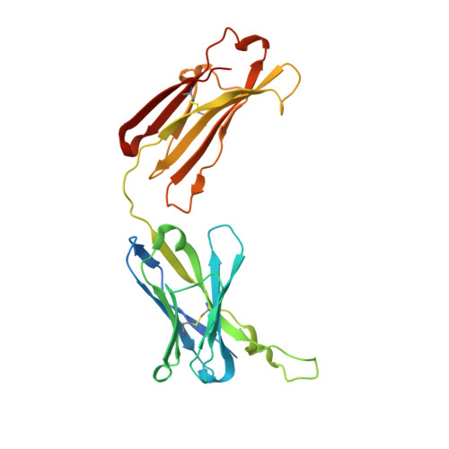
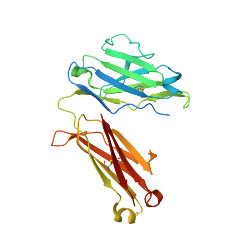
Crystal structure of the Fab region of a neutralizing antibody against granulocyte-macrophage colony-stimulating factor.

Angkawidjaja, C.Torashima, T.

(2019) Acta Crystallogr F Struct Biol Commun 75: 634-639

  • DOI: https://doi.org/10.1107/S2053230X1901238X
  • Primary Citation Related Structures: 
    5ZMJ

  • PubMed Abstract: 

    An increased level of granulocyte-macrophage colony-stimulating factor has a potential role in the development of autoimmune diseases, and the neutralization of its activity by monoclonal antibodies is a promising therapy for some diseases. Here, the crystal structure of the Fab region of EV1007, a fully human antibody expressed in Chinese hamster ovary cells that was developed from human peripheral blood mononuclear cells, is described. The structure closely resembles that of MB007, which is the Fab region of the same antibody expressed in Escherichia coli [Blech et al. (2012), Biochem. J. 447, 205-215], except at the hinge regions between the immunoglobulin domains and the H3 loop region. This paper presents evidence for the flexibility of the hinge and H3 loop regions of the antibody based on the comparison of two independently solved crystal structures.


  • Organizational Affiliation
    • Antibody Drug Discovery Laboratory, Evec Inc., Technopark 1-chome, Sapporo Electronics Center, Sapporo, Hokkaido, Japan.

Macromolecule Content 

  • Total Structure Weight: 52.37 kDa 
  • Atom Count: 3,830 
  • Modeled Residue Count: 438 
  • Deposited Residue Count: 483 
  • Unique protein chains: 2

Macromolecules

Find similar proteins by:|  3D Structure
Entity ID: 1
MoleculeChains  Sequence LengthOrganismDetailsImage
Heavy chain of a Fab fraction of human IgGA [auth H]248Homo sapiensMutation(s): 0 
Find similar proteins by:|  3D Structure
Entity ID: 2
MoleculeChains  Sequence LengthOrganismDetailsImage
Light chain of a Fab fraction of human IgGB [auth L]235Homo sapiensMutation(s): 0 

Small Molecules

Ligands 2 Unique
IDChains Name / Formula / InChI Key2D Diagram3D Interactions
SO4

Query on SO4


Download:Ideal Coordinates CCD File
C [auth H]
F [auth L]
G [auth L]
H [auth L]
I [auth L]
C [auth H],
F [auth L],
G [auth L],
H [auth L],
I [auth L],
J [auth L]
SULFATE ION
O4 S
QAOWNCQODCNURD-UHFFFAOYSA-L
NA

Query on NA


Download:Ideal Coordinates CCD File
D [auth H],
E [auth H],
K [auth L],
L
SODIUM ION
Na
FKNQFGJONOIPTF-UHFFFAOYSA-N

Experimental Data & Validation

Experimental Data

  • Method: X-RAY DIFFRACTION
  • Resolution: 1.81 Å
  • R-Value Free:  0.191 (Depositor), 0.213 (DCC) 
  • R-Value Work:  0.161 (Depositor) 
  • R-Value Observed: 0.163 (Depositor) 
Space Group: P 21 21 21
Unit Cell:
Length ( Å )Angle ( ˚ )
a = 59.12α = 90
b = 66.69β = 90
c = 129.04γ = 90
Software Package:
Software NamePurpose
REFMACrefinement
iMOSFLMdata reduction
pointlessdata scaling
MOLREPphasing

Structure Validation

View Full Validation Report


Entry History 

Deposition Data

Revision History  (Full details and data files)

  • Version 1.0: 2019-04-17
    Type: Initial release
  • Version 1.1: 2019-10-16
    Changes: Data collection, Database references
  • Version 1.2: 2023-11-22
    Changes: Data collection, Database references, Derived calculations, Refinement description
  • Version 1.3: 2024-11-13
    Changes: Structure summary