5ZLP | pdb_00005zlp

Crystal structure of glutamine synthetase from helicobacter pylori


Experimental Data Snapshot

  • Method: X-RAY DIFFRACTION
  • Resolution: 2.93 Å
  • R-Value Free: 
    0.246 (Depositor), 0.245 (DCC) 
  • R-Value Work: 
    0.168 (Depositor), 0.170 (DCC) 
  • R-Value Observed: 
    0.172 (Depositor) 

wwPDB Validation 3D Report Full Report

Validation slider image for 5ZLP

Ligand Structure Quality Assessment 


This is version 1.1 of the entry. See complete history

Literature

Structural Analysis of Glutamine Synthetase from Helicobacter pylori.

Joo, H.K.Park, Y.W.Jang, Y.Y.Lee, J.Y.

(2018) Sci Rep 8: 11657-11657

  • DOI: https://doi.org/10.1038/s41598-018-30191-5
  • Primary Citation Related Structures: 
    5ZLI, 5ZLP

  • PubMed Abstract: 

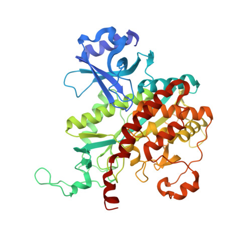
    Glutamine synthetase (GS) is an enzyme that regulates nitrogen metabolism and synthesizes glutamine via glutamate, ATP, and ammonia. GS is a homo-oligomeric protein of eight, ten, or twelve subunits, and each subunit-subunit interface has its own active site. GS can be divided into GS I, GS II, and GS III. GS I and GS III form dodecamer in bacteria and archaea, whereas GS II form decamer in eukaryotes. GS I can be further subdivided into GS I-α and GS I-β according to its sequence and regulatory mechanism. GS is an essential protein for the survival of Helicobacter pylori which its infection could promote gastroduodenal diseases. Here, we determined the crystal structures of the GS from H. pylori (Hpy GS) in its apo- and substrate-bound forms at 2.8 Å and 2.9 Å resolution, respectively. Hpy GS formed a dodecamer composed of two hexameric rings stacked face-to-face. Hpy GS, which belongs to GS I, cannot be clearly classified as either GS I-α or GS I-β based on its sequence and regulatory mechanism. In this study, we propose that Hpy GS could be classified as a new GS-I subfamily and provide structural information on the apo- and substrate-bound forms of the protein.


  • Organizational Affiliation
    • Department of Life Science, Dongguk University-Seoul, Ilsandong-gu, Goyang-si, Gyeonggi-do, 10326, Republic of Korea.

Macromolecule Content 

  • Total Structure Weight: 661.46 kDa 
  • Atom Count: 46,003 
  • Modeled Residue Count: 5,704 
  • Deposited Residue Count: 5,772 
  • Unique protein chains: 1

Macromolecules

Find similar proteins by:|  3D Structure
Entity ID: 1
MoleculeChains  Sequence LengthOrganismDetailsImage
Glutamine synthetase481Helicobacter pylori 26695Mutation(s): 0 
Gene Names: glnAHP_0512
EC: 6.3.1.2
UniProt
Find proteins for P94845 (Helicobacter pylori (strain ATCC 700392 / 26695))
Explore P94845 
Go to UniProtKB:  P94845
Entity Groups
Sequence Clusters30% Identity50% Identity70% Identity90% Identity95% Identity100% Identity
UniProt GroupP94845
Sequence Annotations
Expand
Reference Sequence

Small Molecules

Ligands 5 Unique
IDChains Name / Formula / InChI Key2D Diagram3D Interactions
ATP

Query on ATP


Download:Ideal Coordinates CCD File
CA [auth G]
M [auth A]
N [auth B]
O [auth C]
R [auth F]
CA [auth G],
M [auth A],
N [auth B],
O [auth C],
R [auth F],
W [auth K]
ADENOSINE-5'-TRIPHOSPHATE
C10 H16 N5 O13 P3
ZKHQWZAMYRWXGA-KQYNXXCUSA-N
ADP

Query on ADP


Download:Ideal Coordinates CCD File
AA [auth L]
DA [auth I]
P [auth D]
Q [auth E]
S [auth H]
AA [auth L],
DA [auth I],
P [auth D],
Q [auth E],
S [auth H],
T [auth J]
ADENOSINE-5'-DIPHOSPHATE
C10 H15 N5 O10 P2
XTWYTFMLZFPYCI-KQYNXXCUSA-N
P3P

Query on P3P


Download:Ideal Coordinates CCD File
Y [auth L](2S)-2-AMINO-4-[METHYL(PHOSPHONOOXY)PHOSPHORYL]BUTANOIC ACID
C5 H13 N O7 P2
UXRZHSKRCMUNIN-YZSVAWROSA-N
PPQ

Query on PPQ


Download:Ideal Coordinates CCD File
EA [auth I],
U [auth J],
X [auth K]
PHOSPHINOTHRICIN
C5 H12 N O4 P
IAJOBQBIJHVGMQ-BYPYZUCNSA-N
MG

Query on MG


Download:Ideal Coordinates CCD File
BA [auth L],
FA [auth I],
V [auth J],
Z [auth L]
MAGNESIUM ION
Mg
JLVVSXFLKOJNIY-UHFFFAOYSA-N

Experimental Data & Validation

Experimental Data

  • Method: X-RAY DIFFRACTION
  • Resolution: 2.93 Å
  • R-Value Free:  0.246 (Depositor), 0.245 (DCC) 
  • R-Value Work:  0.168 (Depositor), 0.170 (DCC) 
  • R-Value Observed: 0.172 (Depositor) 
Space Group: C 1 2 1
Unit Cell:
Length ( Å )Angle ( ˚ )
a = 234.409α = 90
b = 135.167β = 91.61
c = 203.08γ = 90
Software Package:
Software NamePurpose
HKL-2000data scaling
PHENIXrefinement
PDB_EXTRACTdata extraction
HKL-2000data reduction
PHASERphasing

Structure Validation

View Full Validation Report


Ligand Structure Quality Assessment 


Entry History 

& Funding Information

Deposition Data


Funding OrganizationLocationGrant Number
National Research Foundation (Korea)Korea, Republic Of2017R1D1A1B03032109
Korea, Republic Of710013-03-1-SB120

Revision History  (Full details and data files)

  • Version 1.0: 2018-08-29
    Type: Initial release
  • Version 1.1: 2024-03-27
    Changes: Data collection, Database references, Derived calculations