5ZJM | pdb_00005zjm

Crystal structure of N-acetylneuraminate lyase from Fusobacterium nucleatum


Experimental Data Snapshot

  • Method: X-RAY DIFFRACTION
  • Resolution: 2.32 Å
  • R-Value Free: 
    0.245 (Depositor), 0.250 (DCC) 
  • R-Value Work: 
    0.202 (Depositor), 0.208 (DCC) 
  • R-Value Observed: 
    0.204 (Depositor) 

Starting Model: experimental
View more details

wwPDB Validation 3D Report Full Report

Validation slider image for 5ZJM

This is version 1.1 of the entry. See complete history

Literature

Crystal structures and kinetics of N-acetylneuraminate lyase from Fusobacterium nucleatum

Kumar, J.P.Rao, H.Nayak, V.Ramaswamy, S.

(2018) Acta Crystallogr F Struct Biol Commun 74: 725-732

  • DOI: https://doi.org/10.1107/S2053230X18012992
  • Primary Citation Related Structures: 
    5ZJM, 5ZKA

  • PubMed Abstract: 

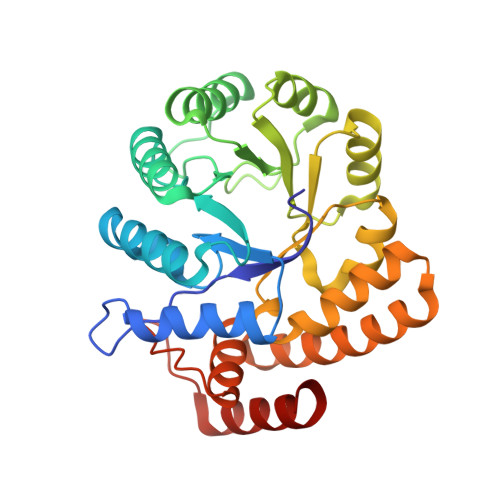
    N-Acetyl-D-neuraminic acid lyase (NanA) catalyzes the breakdown of sialic acid (Neu5Ac) to N-acetyl-D-mannosamine (ManNAc) and pyruvate. NanA plays a key role in Neu5Ac catabolism in many pathogenic and bacterial commensals where sialic acid is available as a carbon and nitrogen source. Several pathogens or commensals decorate their surfaces with sialic acids as a strategy to escape host innate immunity. Catabolism of sialic acid is key to a range of host-pathogen interactions. In this study, atomic resolution structures of NanA from Fusobacterium nucleatum (FnNanA) in ligand-free and ligand-bound forms are reported at 2.32 and 1.76 Å resolution, respectively. F. nucleatum is a Gram-negative pathogen that causes gingival and periodontal diseases in human hosts. Like other bacterial N-acetylneuraminate lyases, FnNanA also shares the triosephosphate isomerase (TIM)-barrel fold. As observed in other homologous enzymes, FnNanA forms a tetramer. In order to characterize the structure-function relationship, the steady-state kinetic parameters of the enzyme are also reported.


  • Organizational Affiliation
    • Technologies for the Advancement of Science, Institute for Stem Cell Biology and Regenerative Medicine, NCBS, GKVK Campus, Bangalore, Karnataka 560 065, India.

Macromolecule Content 

  • Total Structure Weight: 131.92 kDa 
  • Atom Count: 8,907 
  • Modeled Residue Count: 1,162 
  • Deposited Residue Count: 1,164 
  • Unique protein chains: 1

Macromolecules

Find similar proteins by:|  3D Structure
Entity ID: 1
MoleculeChains  Sequence LengthOrganismDetailsImage
N-acetylneuraminate lyase
A, B, C, D
291Fusobacterium nucleatum subsp. nucleatum ATCC 25586Mutation(s): 0 
Gene Names: nanA
EC: 4.1.3.3
UniProt
Find proteins for Q8RDN6 (Fusobacterium nucleatum subsp. nucleatum (strain ATCC 25586 / DSM 15643 / BCRC 10681 / CIP 101130 / JCM 8532 / KCTC 2640 / LMG 13131 / VPI 4355))
Explore Q8RDN6 
Go to UniProtKB:  Q8RDN6
Entity Groups
Sequence Clusters30% Identity50% Identity70% Identity90% Identity95% Identity100% Identity
UniProt GroupQ8RDN6
Sequence Annotations
Expand
Reference Sequence

Experimental Data & Validation

Experimental Data

  • Method: X-RAY DIFFRACTION
  • Resolution: 2.32 Å
  • R-Value Free:  0.245 (Depositor), 0.250 (DCC) 
  • R-Value Work:  0.202 (Depositor), 0.208 (DCC) 
  • R-Value Observed: 0.204 (Depositor) 
Space Group: C 1 2 1
Unit Cell:
Length ( Å )Angle ( ˚ )
a = 105.165α = 90
b = 108.977β = 96.88
c = 141.137γ = 90
Software Package:
Software NamePurpose
PHENIXrefinement
Aimlessdata scaling
PDB_EXTRACTdata extraction
XDSdata reduction
PHENIXphasing

Structure Validation

View Full Validation Report


Entry History 

& Funding Information

Deposition Data


Funding OrganizationLocationGrant Number
IndiaBT/IN/SWEDEN/41/SR/2013
IndiaBT/PR5081/INF/156/2012
IndiaBT/PR12422/MED/31/287/214
IndiaBT/INF/22/SP22660/2017

Revision History  (Full details and data files)

  • Version 1.0: 2019-01-23
    Type: Initial release
  • Version 1.1: 2023-11-22
    Changes: Data collection, Database references, Refinement description