5ZFP

Structure of the ExbB/ExbD hexameric complex


Experimental Data Snapshot

  • Method: X-RAY DIFFRACTION
  • Resolution: 2.84 Å
  • R-Value Free: 0.288 
  • R-Value Work: 0.245 
  • R-Value Observed: 0.247 

wwPDB Validation   3D Report Full Report


This is version 1.1 of the entry. See complete history


Literature

Hexameric and pentameric complexes of the ExbBD energizer in the Ton system.

Maki-Yonekura, S.Matsuoka, R.Yamashita, Y.Shimizu, H.Tanaka, M.Iwabuki, F.Yonekura, K.

(2018) Elife 7

  • DOI: https://doi.org/10.7554/eLife.35419
  • Primary Citation of Related Structures:  
    5ZFP, 5ZFU, 5ZFV

  • PubMed Abstract: 

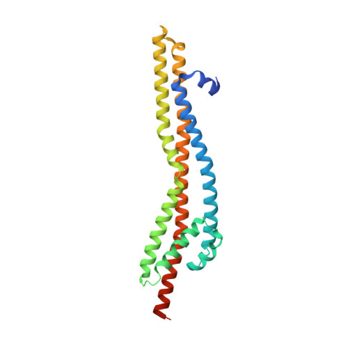
    Gram-negative bacteria import essential nutrients such as iron and vitamin B 12 through outer membrane receptors. This process utilizes proton motive force harvested by the Ton system made up of three inner membrane proteins, ExbB, ExbD and TonB. ExbB and ExbD form the proton channel that energizes uptake through TonB. Recently, crystal structures suggest that the ExbB pentamer is the scaffold. Here, we present structures of hexameric complexes of ExbB and ExbD revealed by X-ray crystallography and single particle cryo-EM. Image analysis shows that hexameric and pentameric complexes coexist, with the proportion of hexamer increasing with pH. Channel current measurement and 2D crystallography support the existence and transition of the two oligomeric states in membranes. The hexameric complex consists of six ExbB subunits and three ExbD transmembrane helices enclosed within the central channel. We propose models for activation/inactivation associated with hexamer and pentamer formation and utilization of proton motive force.


  • Organizational Affiliation

    Biostructural Mechanism Laboratory, RIKEN SPring-8 Center, Sayo, Japan.


Macromolecules
Find similar proteins by:  (by identity cutoff)  |  3D Structure
Entity ID: 1
MoleculeChains Sequence LengthOrganismDetailsImage
Biopolymer transport protein ExbB
A, B, C, D, E
A, B, C, D, E, F, G, H, I, J, K, L
244Escherichia coli K-12Mutation(s): 0 
Gene Names: exbBb3006JW2974
Membrane Entity: Yes 
UniProt
Find proteins for P0ABU7 (Escherichia coli (strain K12))
Explore P0ABU7 
Go to UniProtKB:  P0ABU7
Entity Groups  
Sequence Clusters30% Identity50% Identity70% Identity90% Identity95% Identity100% Identity
UniProt GroupP0ABU7
Sequence Annotations
Expand
  • Reference Sequence
Experimental Data & Validation

Experimental Data

  • Method: X-RAY DIFFRACTION
  • Resolution: 2.84 Å
  • R-Value Free: 0.288 
  • R-Value Work: 0.245 
  • R-Value Observed: 0.247 
  • Space Group: P 1 21 1
Unit Cell:
Length ( Å )Angle ( ˚ )
a = 121.47α = 90
b = 106.34β = 111.75
c = 163.36γ = 90
Software Package:
Software NamePurpose
BUSTERrefinement
XDSdata reduction
XDSdata scaling
PHASERphasing

Structure Validation

View Full Validation Report


Entry History & Funding Information

Deposition Data


Funding OrganizationLocationGrant Number
the Japan Society for the Promotion of Science Grant-in-Aid for Scientific Research GrantJapan15K06986
the Japan Society for the Promotion of Science Grant-in-Aid for Scientific Research GrantJapan16H04757

Revision History  (Full details and data files)

  • Version 1.0: 2018-05-09
    Type: Initial release
  • Version 1.1: 2023-11-22
    Changes: Data collection, Database references, Refinement description