


Crystal Structure of RNF13 RING domain
Sarkar, S., Datta, A.B.To be published.
Experimental Data Snapshot
wwPDB Validation 3D Report Full Report

Entity ID: 1 | |||||
|---|---|---|---|---|---|
| Molecule | Chains | Sequence Length | Organism | Details | Image |
| E3 ubiquitin-protein ligase RNF13 | A, B [auth D] | 76 | Homo sapiens | Mutation(s): 0 Gene Names: RNF13, RZF EC: 2.3.2.27 | 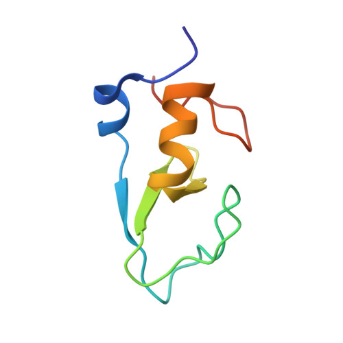 |
UniProt & NIH Common Fund Data Resources | |||||
PHAROS: O43567 GTEx: ENSG00000082996 | |||||
Entity Groups | |||||
| Sequence Clusters | 30% Identity50% Identity70% Identity90% Identity95% Identity100% Identity | ||||
| UniProt Group | O43567 | ||||
Sequence AnnotationsExpand | |||||
Reference Sequence | |||||
| Ligands 1 Unique | |||||
|---|---|---|---|---|---|
| ID | Chains | Name / Formula / InChI Key | 2D Diagram | 3D Interactions | |
| ZN Download:Ideal Coordinates CCD File | C [auth A], D [auth A], E [auth D], F [auth D] | ZINC ION Zn PTFCDOFLOPIGGS-UHFFFAOYSA-N |  | ||
| Length ( Å ) | Angle ( ˚ ) |
|---|---|
| a = 90.338 | α = 90 |
| b = 90.338 | β = 90 |
| c = 45.445 | γ = 120 |
| Software Name | Purpose |
|---|---|
| PHENIX | refinement |
| XDS | data reduction |
| Aimless | data scaling |
| SHELXD | phasing |
| Funding Organization | Location | Grant Number |
|---|---|---|
| Wellcome Trust | India | 500241/Z/11/Z |


