5Z1B | pdb_00005z1b

Structure of Bifidobacterium dentium beta-glucuronidase complexed with coumarin-3-O-glucuronide


Experimental Data Snapshot

  • Method: X-RAY DIFFRACTION
  • Resolution: 1.45 Å
  • R-Value Free: 
    0.160 (Depositor), 0.162 (DCC) 
  • R-Value Work: 
    0.130 (Depositor), 0.136 (DCC) 
  • R-Value Observed: 
    0.130 (Depositor) 

wwPDB Validation 3D Report Full Report

Validation slider image for 5Z1B

Ligand Structure Quality Assessment 


This is version 1.2 of the entry. See complete history

Literature

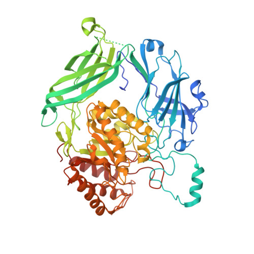
Dissection of the substrate preference and structure of gut microbial-glucuronidases identifies the major bacteria causing xenobiotic toxicity

Dashnyam, P.Mudududdla, R.Hsieh, T.J.Lin, T.C.Lin, H.Y.Chen, P.Y.Hsu, C.Y.Lin, C.H.

To be published.

Macromolecule Content 

  • Total Structure Weight: 309.95 kDa 
  • Atom Count: 22,105 
  • Modeled Residue Count: 2,493 
  • Deposited Residue Count: 2,768 
  • Unique protein chains: 1

Macromolecules

Find similar proteins by:|  3D Structure
Entity ID: 1
MoleculeChains  Sequence LengthOrganismDetailsImage
Glycosyl hydrolase family 2, TIM barrel domain protein
A, B, C, D
692Bifidobacterium dentium ATCC 27679Mutation(s): 1 
Gene Names: uidAHMPREF0168_2111
EC: 3.2.1.31
UniProt
Find proteins for E0QAF3 (Bifidobacterium dentium ATCC 27679)
Explore E0QAF3 
Go to UniProtKB:  E0QAF3
Entity Groups
Sequence Clusters30% Identity50% Identity70% Identity90% Identity95% Identity100% Identity
UniProt GroupE0QAF3
Sequence Annotations
Expand
Reference Sequence

Small Molecules

Ligands 2 Unique
IDChains Name / Formula / InChI Key2D Diagram3D Interactions
BDP

Query on BDP


Download:Ideal Coordinates CCD File
E [auth A],
G [auth B],
I [auth C],
K [auth D]
beta-D-glucopyranuronic acid
C6 H10 O7
AEMOLEFTQBMNLQ-QIUUJYRFSA-N
SE2

Query on SE2


Download:Ideal Coordinates CCD File
F [auth A],
H [auth B],
J [auth C],
L [auth D]
3-HYDROXY-2H-CHROMEN-2-ONE
C9 H6 O3
MJKVTPMWOKAVMS-UHFFFAOYSA-N

Experimental Data & Validation

Experimental Data

  • Method: X-RAY DIFFRACTION
  • Resolution: 1.45 Å
  • R-Value Free:  0.160 (Depositor), 0.162 (DCC) 
  • R-Value Work:  0.130 (Depositor), 0.136 (DCC) 
  • R-Value Observed: 0.130 (Depositor) 
Space Group: P 1 21 1
Unit Cell:
Length ( Å )Angle ( ˚ )
a = 91.34α = 90
b = 104.165β = 90.4
c = 160.673γ = 90
Software Package:
Software NamePurpose
HKL-2000data scaling
PHENIXrefinement
PDB_EXTRACTdata extraction
HKL-2000data reduction
PHENIXphasing

Structure Validation

View Full Validation Report


Ligand Structure Quality Assessment 


Entry History 

Deposition Data

Revision History  (Full details and data files)

  • Version 1.0: 2018-12-12
    Type: Initial release
  • Version 1.1: 2020-07-29
    Type: Remediation
    Reason: Carbohydrate remediation
    Changes: Data collection, Derived calculations, Structure summary
  • Version 1.2: 2024-03-27
    Changes: Data collection, Database references, Refinement description, Structure summary