Improvement of ST0452N-Acetylglucosamine-1-Phosphate Uridyltransferase Activity by the Cooperative Effect of Two Single Mutations Identified through Structure-Based Protein Engineering
Honda, Y., Nakano, S., Ito, S., Dadashipour, M., Zhang, Z., Kawarabayasi, Y.(2018) Appl Environ Microbiol 84
- PubMed: 30291121 Search on PubMedSearch on PubMed Central
- DOI: https://doi.org/10.1128/AEM.02213-18
- Primary Citation Related Structures:
5Z09, 5Z0A - PubMed Abstract:
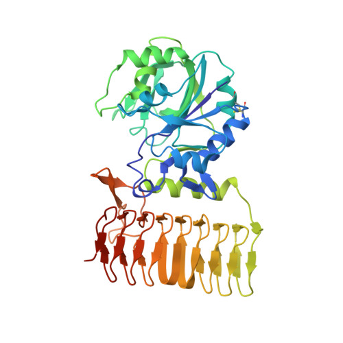
We showed previously that the Y97N mutant of the ST0452 protein, isolated from Sulfolobus tokodaii , exhibited over 4 times higher N -acetylglucosamine-1-phosphate (GlcNAc-1-P) uridyltransferase (UTase) activity, compared with that of the wild-type ST0452 protein. We determined the three-dimensional structure of the Y97N protein to explore the detailed mechanism underlying this increased activity. The overall structure was almost identical to that of the wild-type ST0452 protein (PDB ID 2GGO), with residue 97 (Asn) interacting with the O-5 atom of N -acetylglucosamine (GlcNAc) in the complex without metal ions. The same interaction was observed for Escherichia coli GlmU in the absence of metal ions. These observations indicated that the three-dimensional structure of the Y97N protein was not changed by this substitution but the interactions with the substrate were slightly modified, which might cause the activity to increase. The crystal structure of the Y97N protein also showed that positions 146 (Glu) and 80 (Thr) formed interactions with GlcNAc, and an engineering strategy was applied to these residues to increase activity. All proteins substituted at position 146 had drastically decreased activities, whereas several proteins substituted at position 80 showed higher GlcNAc-1-P UTase activity, compared to that of the wild-type protein. The substituted amino acids at positions 80 and 97 might result in optimized interactions with the substrate; therefore, we predicted that the combination of these two substitutions might cooperatively increase GlcNAc-1-P UTase activity. Of the four double mutant ST0452 proteins generated, T80S/Y97N showed 6.5-times-higher activity, compared to that of the wild-type ST0452 protein, revealing that these two substituted residues functioned cooperatively to increase GlcNAc-1-P UTase activity. IMPORTANCE We demonstrated that the enzymatic activity of a thermostable protein was over 4 times higher than that of the wild-type protein following substitution of a single amino acid, without affecting its thermostability. The three-dimensional structure of the improved mutant protein complexed with substrate was determined. The same overall structure and interaction between the substituted residue and the GlcNAc substrate as observed in the well-characterized bacterial enzyme suggested that the substitution of Tyr at position 97 by Asn might slightly change the interaction. This subtle change in the interaction might potentially increase the GlcNAc-1-P UTase activity of the mutant protein. These observations indicated that a drastic change in the structure of a natural thermostable enzyme is not necessary to increase its activity; a subtle change in the interaction with the substrate might be sufficient. Cooperative effects were observed in the appropriate double mutant protein. This work provides useful information for the future engineering of natural enzymes.
- Laboratory for Functional Genomics of Extremophiles, Faculty of Agriculture, Kyushu University, Fukuoka, Japan.
Organizational Affiliation: