5YY8 | pdb_00005yy8

Crystal structure of the Kelch domain of human NS1-BP


Experimental Data Snapshot

  • Method: X-RAY DIFFRACTION
  • Resolution: 1.98 Å
  • R-Value Free: 
    0.232 (Depositor), 0.230 (DCC) 
  • R-Value Work: 
    0.189 (Depositor), 0.189 (DCC) 
  • R-Value Observed: 
    0.192 (Depositor) 

wwPDB Validation 3D Report Full Report

Validation slider image for 5YY8

This is version 1.1 of the entry. See complete history

Literature

Crystal structure of the Kelch domain of human NS1-binding protein at 1.98 angstrom resolution.

Guo, L.Liu, Y.

(2018) Acta Crystallogr F Struct Biol Commun 74: 174-178

  • DOI: https://doi.org/10.1107/S2053230X18001577
  • Primary Citation Related Structures: 
    5YY8

  • PubMed Abstract: 

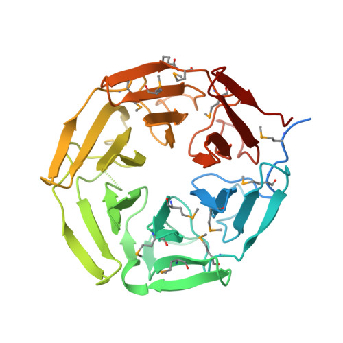
    NS1-binding protein (NS1-BP), which belongs to the Kelch protein superfamily, was first identified as a novel human 70 kDa protein that interacts with NS1 of Influenza A virus. It is involved in many cell functions, including pre-mRNA splicing, the ERK signalling pathway, the aryl hydrocarbon receptor (AHR) pathway, F-actin organization and protein ubiquitylation. However, the structure of NS1-BP is still unknown, which may impede functional studies. Here, the structure of the C-terminal Kelch domain of NS1-BP (NS1-BP-C; residues 330-642) was determined at 1.98 Å resolution. The Kelch domain adopts a highly symmetric six-bladed β-propeller fold structure. Each blade of the β-propeller is composed of four antiparallel β-strands. Comparison of the Kelch-domain structures of NS1-BP and its homologues showed that the Gly-Gly pair in β-strand B and the hydrophobic Trp residue in β-strand D are highly conserved, while the B-C loops in blades 2 and 6 are variable. This structure of the Kelch domain of NS1-BP extends the understanding of NS1-BP.


  • Organizational Affiliation
    • National Laboratory of Biomacromolecules, Institute of Biophysics, Chinese Academy of Sciences, Beijing 100101, People's Republic of China.

Macromolecule Content 

  • Total Structure Weight: 37.03 kDa 
  • Atom Count: 2,314 
  • Modeled Residue Count: 284 
  • Deposited Residue Count: 328 
  • Unique protein chains: 1

Macromolecules

Find similar proteins by:|  3D Structure
Entity ID: 1
MoleculeChains  Sequence LengthOrganismDetailsImage
Influenza virus NS1A-binding protein328Homo sapiensMutation(s): 0 
Gene Names: IVNS1ABPARA3FLARA3KIAA0850NS1NS1BPHSPC068
UniProt & NIH Common Fund Data Resources
Find proteins for Q9Y6Y0 (Homo sapiens)
Explore Q9Y6Y0 
Go to UniProtKB:  Q9Y6Y0
PHAROS:  Q9Y6Y0
GTEx:  ENSG00000116679 
Entity Groups
Sequence Clusters30% Identity50% Identity70% Identity90% Identity95% Identity100% Identity
UniProt GroupQ9Y6Y0
Sequence Annotations
Expand
Reference Sequence

Small Molecules

Modified Residues  1 Unique
IDChains TypeFormula2D DiagramParent
MSE
Query on MSE
A
L-PEPTIDE LINKINGC5 H11 N O2 SeMET

Experimental Data & Validation

Experimental Data

  • Method: X-RAY DIFFRACTION
  • Resolution: 1.98 Å
  • R-Value Free:  0.232 (Depositor), 0.230 (DCC) 
  • R-Value Work:  0.189 (Depositor), 0.189 (DCC) 
  • R-Value Observed: 0.192 (Depositor) 
Space Group: P 32 2 1
Unit Cell:
Length ( Å )Angle ( ˚ )
a = 61.556α = 90
b = 61.556β = 90
c = 140.572γ = 120
Software Package:
Software NamePurpose
PHENIXrefinement
HKL-2000data reduction
HKL-2000data scaling

Structure Validation

View Full Validation Report


Entry History 

Deposition Data

Revision History  (Full details and data files)

  • Version 1.0: 2018-03-14
    Type: Initial release
  • Version 1.1: 2024-10-16
    Changes: Data collection, Database references, Structure summary