5YUN | pdb_00005yun

Crystal structure of SSB complexed with myc


Experimental Data Snapshot

  • Method: X-RAY DIFFRACTION
  • Resolution: 2.67 Å
  • R-Value Free: 
    0.256 (Depositor), 0.263 (DCC) 
  • R-Value Work: 
    0.207 (Depositor), 0.215 (DCC) 
  • R-Value Observed: 
    0.210 (Depositor) 

Starting Model: experimental
View more details

wwPDB Validation 3D Report Full Report

Validation slider image for 5YUN

Ligand Structure Quality Assessment 


This is version 1.2 of the entry. See complete history

Literature

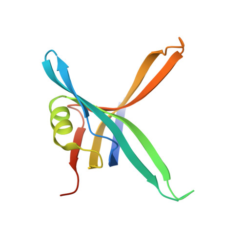
Crystal structure of SSB complexed with inhibitor myricetin.

Huang, C.Y.

(2018) Biochem Biophys Res Commun 504: 704-708

  • DOI: https://doi.org/10.1016/j.bbrc.2018.08.188
  • Primary Citation Related Structures: 
    5YUN

  • PubMed Abstract: 

    Single-stranded DNA-binding protein (SSB) is essential for all DNA-dependent cellular processes. SSB inhibitors have been recently suggested as broad-spectrum antibacterial agents in antibiotic development. In this paper, we report the first inhibitor-complexed crystal structure of SSB from Pseudomonas aeruginosa PAO1 (PaSSB) at 2.68 Å resolution (PDB entry 5YUN). The inhibitor, myricetin, is a flavonol that possesses many pharmacological activities, such as anticancer, anti-inflammatory, and antibacterial properties, and is beneficial for humans. Four monomers of PaSSB and two of myricetins were found per asymmetric unit. Various interactions between myricetin and PaSSB were examined. Among these, four residues in PaSSB, Lys7, Arg62, Glu80, and Gly107 were found crucial for forming hydrogen bond to myricetin. These two myricetins occupy the grooves for ssDNA-binding of SSB that may prevent ssDNA-wrapping and ssDNA-binding stably from SSB. In addition to explaining how SSB can be inhibited, the myricetin-SSB interaction modes in this paper may also provide insights into how myricetin can bind and inhibit proteins on cancer-signaling pathways.


  • Organizational Affiliation
    • School of Biomedical Sciences, Chung Shan Medical University, No.110, Sec.1, Chien-Kuo N. Rd., Taichung City, Taiwan; Department of Medical Research, Chung Shan Medical University Hospital, No.110, Sec.1, Chien-Kuo N. Rd, Taichung City, Taiwan. Electronic address: cyhuang@csmu.edu.tw.

Macromolecule Content 

  • Total Structure Weight: 55.73 kDa 
  • Atom Count: 3,310 
  • Modeled Residue Count: 411 
  • Deposited Residue Count: 484 
  • Unique protein chains: 1

Macromolecules

Find similar proteins by:|  3D Structure
Entity ID: 1
MoleculeChains  Sequence LengthOrganismDetailsImage
Single-stranded DNA-binding protein
A, B, C, D
121Pseudomonas aeruginosa PAO1Mutation(s): 0 
Gene Names: ssbPA4232
UniProt
Find proteins for P40947 (Pseudomonas aeruginosa (strain ATCC 15692 / DSM 22644 / CIP 104116 / JCM 14847 / LMG 12228 / 1C / PRS 101 / PAO1))
Explore P40947 
Go to UniProtKB:  P40947
Entity Groups
Sequence Clusters30% Identity50% Identity70% Identity90% Identity95% Identity100% Identity
UniProt GroupP40947
Sequence Annotations
Expand
Reference Sequence

Small Molecules

Experimental Data & Validation

Experimental Data

  • Method: X-RAY DIFFRACTION
  • Resolution: 2.67 Å
  • R-Value Free:  0.256 (Depositor), 0.263 (DCC) 
  • R-Value Work:  0.207 (Depositor), 0.215 (DCC) 
  • R-Value Observed: 0.210 (Depositor) 
Space Group: P 31
Unit Cell:
Length ( Å )Angle ( ˚ )
a = 60.11α = 90
b = 60.11β = 90
c = 130.868γ = 120
Software Package:
Software NamePurpose
REFMACrefinement
HKL-2000data reduction
HKL-2000data scaling
MOLREPphasing

Structure Validation

View Full Validation Report


Ligand Structure Quality Assessment 


Entry History 

Deposition Data

Revision History  (Full details and data files)

  • Version 1.0: 2018-10-10
    Type: Initial release
  • Version 1.1: 2018-10-17
    Changes: Data collection, Database references, Structure summary
  • Version 1.2: 2023-11-22
    Changes: Data collection, Database references, Refinement description