5YSE | pdb_00005yse

Crystal structure of beta-1,2-glucooligosaccharide binding protein in complex with sophorotetraose


Experimental Data Snapshot

  • Method: X-RAY DIFFRACTION
  • Resolution: 1.60 Å
  • R-Value Free: 
    0.188 (Depositor), 0.227 (DCC) 
  • R-Value Work: 
    0.158 (Depositor), 0.204 (DCC) 
  • R-Value Observed: 
    0.159 (Depositor) 

Starting Model: experimental
View more details

wwPDB Validation 3D Report Full Report

Validation slider image for 5YSE

This is version 2.1 of the entry. See complete history

Literature

Structural and thermodynamic insights into beta-1,2-glucooligosaccharide capture by a solute-binding protein inListeria innocua.

Abe, K.Sunagawa, N.Terada, T.Takahashi, Y.Arakawa, T.Igarashi, K.Samejima, M.Nakai, H.Taguchi, H.Nakajima, M.Fushinobu, S.

(2018) J Biological Chem 293: 8812-8828

  • DOI: https://doi.org/10.1074/jbc.RA117.001536
  • Primary Citation Related Structures: 
    5YSB, 5YSD, 5YSE, 5YSF

  • PubMed Abstract: 

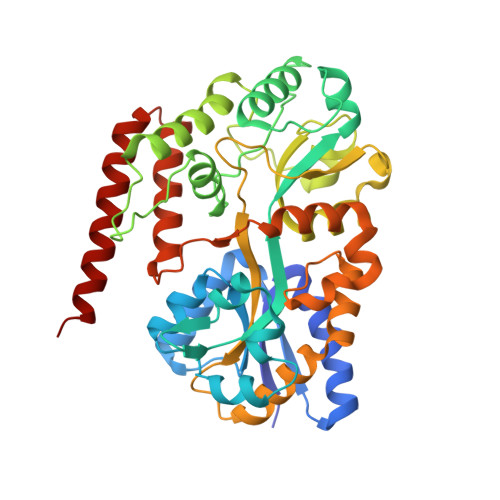
    β-1,2-Glucans are bacterial carbohydrates that exist in cyclic or linear forms and play an important role in infections and symbioses involving Gram-negative bacteria. Although several β-1,2-glucan-associated enzymes have been characterized, little is known about how β-1,2-glucan and its shorter oligosaccharides (Sop n s) are captured and imported into the bacterial cell. Here, we report the biochemical and structural characteristics of the Sop n -binding protein (SO-BP, Lin1841) associated with the ATP-binding cassette (ABC) transporter from the Gram-positive bacterium Listeria innocua Calorimetric analysis revealed that SO-BP specifically binds to Sop n s with a degree of polymerization of 3 or more, with K d values in the micromolar range. The crystal structures of SO-BP in an unliganded open form and in closed complexes with tri-, tetra-, and pentaoligosaccharides (Sop 3-5 ) were determined to a maximum resolution of 1.6 Å. The binding site displayed shape complementarity to Sop n , which adopted a zigzag conformation. We noted that water-mediated hydrogen bonds and stacking interactions play a pivotal role in the recognition of Sop 3-5 by SO-BP, consistent with its binding thermodynamics. Computational free-energy calculations and a mutational analysis confirmed that interactions with the third glucose moiety of Sop n s are significantly responsible for ligand binding. A reduction in unfavorable changes in binding entropy that were in proportion to the lengths of the Sop n s was explained by conformational entropy changes. Phylogenetic and sequence analyses indicated that SO-BP ABC transporter homologs, glycoside hydrolases, and other related proteins are co-localized in the genomes of several bacteria. This study may improve our understanding of bacterial β-1,2-glucan metabolism and promote the discovery of unidentified β-1,2-glucan-associated proteins.


  • Organizational Affiliation
    • From the Department of Biotechnology.

Macromolecule Content 

  • Total Structure Weight: 90.88 kDa 
  • Atom Count: 6,899 
  • Modeled Residue Count: 774 
  • Deposited Residue Count: 794 
  • Unique protein chains: 1

Macromolecules

Find similar proteins by:|  3D Structure
Entity ID: 1
MoleculeChains  Sequence LengthOrganismDetailsImage
Lin1841 protein
A, B
397Listeria innocua Clip11262Mutation(s): 0 
Gene Names: lin1841
UniProt
Find proteins for Q92AS8 (Listeria innocua serovar 6a (strain ATCC BAA-680 / CLIP 11262))
Explore Q92AS8 
Go to UniProtKB:  Q92AS8
Entity Groups
Sequence Clusters30% Identity50% Identity70% Identity90% Identity95% Identity100% Identity
UniProt GroupQ92AS8
Sequence Annotations
Expand
Reference Sequence

Oligosaccharides

Help  
Entity ID: 2
MoleculeChains Length2D Diagram GlycosylationD Interactions
beta-D-glucopyranose-(1-2)-beta-D-glucopyranose-(1-2)-beta-D-glucopyranose-(1-2)-alpha-D-glucopyranose
C, D
4N/A
Glycosylation Resources
GlyTouCan: G06375IT
GlyCosmos: G06375IT
GlyGen: G06375IT

Experimental Data & Validation

Experimental Data

  • Method: X-RAY DIFFRACTION
  • Resolution: 1.60 Å
  • R-Value Free:  0.188 (Depositor), 0.227 (DCC) 
  • R-Value Work:  0.158 (Depositor), 0.204 (DCC) 
  • R-Value Observed: 0.159 (Depositor) 
Space Group: P 1
Unit Cell:
Length ( Å )Angle ( ˚ )
a = 36.014α = 89.99
b = 63.28β = 82.24
c = 87.451γ = 86.08
Software Package:
Software NamePurpose
REFMACrefinement
HKL-2000data processing
MOLREPphasing
ARP/wARPmodel building

Structure Validation

View Full Validation Report


Entry History 

Deposition Data

Revision History  (Full details and data files)

  • Version 1.0: 2018-05-02
    Type: Initial release
  • Version 1.1: 2018-06-20
    Changes: Data collection, Database references
  • Version 2.0: 2020-07-29
    Type: Remediation
    Reason: Carbohydrate remediation
    Changes: Atomic model, Data collection, Derived calculations, Structure summary
  • Version 2.1: 2023-11-22
    Changes: Data collection, Database references, Derived calculations, Refinement description, Structure summary