5YRN

Structure of RIP2 CARD domain


Experimental Data Snapshot

  • Method: ELECTRON MICROSCOPY
  • Resolution: 4.10 Å
  • Aggregation State: FILAMENT 
  • Reconstruction Method: HELICAL 

wwPDB Validation   3D Report Full Report


This is version 1.3 of the entry. See complete history


Literature

Structural basis of RIP2 activation and signaling.

Gong, Q.Long, Z.Zhong, F.L.Teo, D.E.T.Jin, Y.Yin, Z.Boo, Z.Z.Zhang, Y.Zhang, J.Yang, R.Bhushan, S.Reversade, B.Li, Z.Wu, B.

(2018) Nat Commun 9: 4993-4993

  • DOI: https://doi.org/10.1038/s41467-018-07447-9
  • Primary Citation of Related Structures:  
    5YRN

  • PubMed Abstract: 

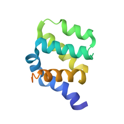
    Signals arising from bacterial infections are detected by pathogen recognition receptors (PRRs) and are transduced by specialized adapter proteins in mammalian cells. The Receptor-interacting-serine/threonine-protein kinase 2 (RIPK2 or RIP2) is such an adapter protein that is critical for signal propagation of the Nucleotide-binding-oligomerization-domain-containing proteins 1/2 (NOD1 and NOD2). Dysregulation of this signaling pathway leads to defects in bacterial detection and in some cases autoimmune diseases. Here, we show that the Caspase-activation-and-recruitment-domain (CARD) of RIP2 (RIP2-CARD) forms oligomeric structures upon stimulation by either NOD1-CARD or NOD2-2CARD. We reconstitute this complex, termed the RIPosome in vitro and solve the cryo-EM filament structure of the active RIP2-CARD complex at 4.1 Å resolution. The structure suggests potential mechanisms by which CARD domains from NOD1 and NOD2 initiate the oligomerization process of RIP2-CARD. Together with structure guided mutagenesis experiments at the CARD-CARD interfaces, we demonstrate molecular mechanisms how RIP2 is activated and self-propagating such signal.


  • Organizational Affiliation

    School of Biological Sciences, Nanyang Technological University, Singapore, 637551, Singapore.


Macromolecules
Find similar proteins by:  (by identity cutoff)  |  3D Structure
Entity ID: 1
MoleculeChains Sequence LengthOrganismDetailsImage
Receptor-interacting serine/threonine-protein kinase 2
A, B, C, D, E
A, B, C, D, E, F, G, H, I, J, K, L
111Homo sapiensMutation(s): 0 
Gene Names: RIPK2CARDIAKRICKRIP2UNQ277/PRO314/PRO34092
EC: 2.7.11.1 (PDB Primary Data), 2.7.10.2 (PDB Primary Data)
UniProt & NIH Common Fund Data Resources
Find proteins for O43353 (Homo sapiens)
Explore O43353 
Go to UniProtKB:  O43353
PHAROS:  O43353
GTEx:  ENSG00000104312 
Entity Groups  
Sequence Clusters30% Identity50% Identity70% Identity90% Identity95% Identity100% Identity
UniProt GroupO43353
Sequence Annotations
Expand
  • Reference Sequence
Experimental Data & Validation

Experimental Data

  • Method: ELECTRON MICROSCOPY
  • Resolution: 4.10 Å
  • Aggregation State: FILAMENT 
  • Reconstruction Method: HELICAL 
EM Software:
TaskSoftware PackageVersion
RECONSTRUCTIONRELION2.0beta
MODEL REFINEMENTCoot0.8.2

Structure Validation

View Full Validation Report


Entry History & Funding Information

Deposition Data


Funding OrganizationLocationGrant Number
Ministry of Education (Singapore)SingaporeNAP SUG

Revision History  (Full details and data files)

  • Version 1.0: 2018-11-14
    Type: Initial release
  • Version 1.1: 2019-05-29
    Changes: Data collection, Database references
  • Version 1.2: 2019-11-06
    Changes: Data collection, Other
  • Version 1.3: 2024-03-27
    Changes: Data collection, Database references