5YQW

Structure and function of a novel periplasmic chitooligosaccharide-binding protein from marine Vibrio bacteria


Experimental Data Snapshot

  • Method: X-RAY DIFFRACTION
  • Resolution: 1.36 Å
  • R-Value Free: 0.226 
  • R-Value Work: 0.195 
  • R-Value Observed: 0.197 

wwPDB Validation   3D Report Full Report


Ligand Structure Quality Assessment 


This is version 2.1 of the entry. See complete history


Literature

Structure and function of a novel periplasmic chitooligosaccharide-binding protein from marineVibriobacteria.

Suginta, W.Sritho, N.Ranok, A.Bulmer, D.M.Kitaoku, Y.van den Berg, B.Fukamizo, T.

(2018) J Biol Chem 293: 5150-5159

  • DOI: https://doi.org/10.1074/jbc.RA117.001012
  • Primary Citation of Related Structures:  
    5YQW

  • PubMed Abstract: 

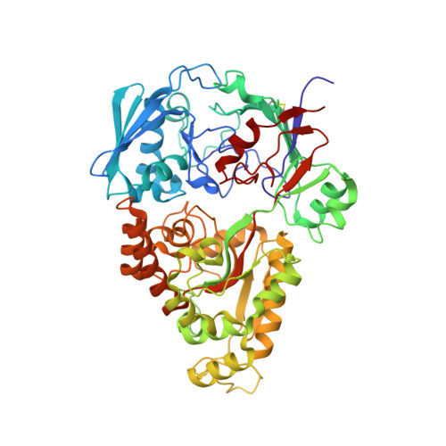
    Periplasmic solute-binding proteins in bacteria are involved in the active transport of nutrients into the cytoplasm. In marine bacteria of the genus Vibrio , a chitooligosaccharide-binding protein (CBP) is thought to be the major solute-binding protein controlling the rate of chitin uptake in these bacteria. However, the molecular mechanism of the CBP involvement in chitin metabolism has not been elucidated. Here, we report the structure and function of a recombinant chitooligosaccharide-binding protein from Vibrio harveyi , namely Vh CBP, expressed in Escherichia coli Isothermal titration calorimetry revealed that Vh CBP strongly binds shorter chitooligosaccharides ((GlcNAc) n , where n = 2, 3, and 4) with affinities that are considerably greater than those for glycoside hydrolase family 18 and 19 chitinases but does not bind longer ones, including insoluble chitin polysaccharides. We also found that VhCBP comprises two domains with flexible linkers and that the domain-domain interface forms the sugar-binding cleft, which is not long extended but forms a small cavity. (GlcNAc) 2 bound to this cavity, apparently triggering a closed conformation of Vh CBP. Trp-363 and Trp-513, which stack against the two individual GlcNAc rings, likely make a major contribution to the high affinity of Vh CBP for (GlcNAc) 2 The strong chitobiose binding, followed by the conformational change of Vh CBP, may facilitate its interaction with an active-transport system in the inner membrane of Vibrio species.


  • Organizational Affiliation

    From the Biochemistry-Electrochemistry Research Unit, Institute of Science, Suranaree University of Technology, Nakhon Ratchasima 30000, Thailand, wipa@sut.ac.th.


Macromolecules
Find similar proteins by:  (by identity cutoff)  |  3D Structure
Entity ID: 1
MoleculeChains Sequence LengthOrganismDetailsImage
Peptide ABC transporter, periplasmic peptide-binding protein532Vibrio harveyi 1DA3Mutation(s): 0 
Gene Names: VME_26970
Entity Groups  
Sequence Clusters30% Identity50% Identity70% Identity90% Identity95% Identity100% Identity
Sequence Annotations
Expand
  • Reference Sequence
Oligosaccharides

Help

Entity ID: 2
MoleculeChains Length2D Diagram Glycosylation3D Interactions
2-acetamido-2-deoxy-beta-D-glucopyranose-(1-4)-2-acetamido-2-deoxy-beta-D-glucopyranose
B
2N/A
Glycosylation Resources
GlyTouCan:  G42666HT
GlyCosmos:  G42666HT
GlyGen:  G42666HT
Experimental Data & Validation

Experimental Data

  • Method: X-RAY DIFFRACTION
  • Resolution: 1.36 Å
  • R-Value Free: 0.226 
  • R-Value Work: 0.195 
  • R-Value Observed: 0.197 
  • Space Group: P 31 2 1
Unit Cell:
Length ( Å )Angle ( ˚ )
a = 54.73α = 90
b = 54.73β = 90
c = 306.442γ = 120
Software Package:
Software NamePurpose
REFMACrefinement
DIALSdata reduction
Aimlessdata scaling
MOLREPphasing

Structure Validation

View Full Validation Report


Ligand Structure Quality Assessment 


Entry History & Funding Information

Deposition Data


Funding OrganizationLocationGrant Number
Office of the Higher Education Commission under the National Research University (NRU) Project of ThailandThailandFtR.12/2558
a Basic Research Grant, Suranaree University of TechnologyThailandBRG578001
SUT grant, Suranaree University of TechnologyThailandSUT1-102-58-36-10

Revision History  (Full details and data files)

  • Version 1.0: 2018-02-21
    Type: Initial release
  • Version 1.1: 2018-04-18
    Changes: Data collection, Database references
  • Version 2.0: 2020-07-29
    Type: Remediation
    Reason: Carbohydrate remediation
    Changes: Atomic model, Data collection, Derived calculations, Non-polymer description, Structure summary
  • Version 2.1: 2023-11-22
    Changes: Data collection, Database references, Refinement description, Structure summary