5YJW

Structure of the Ndi1 protein from Saccharomyces cerevisiae in complex with the competitive inhibitor, stigmatellin.


Experimental Data Snapshot

  • Method: X-RAY DIFFRACTION
  • Resolution: 1.85 Å
  • R-Value Free: 0.204 
  • R-Value Work: 0.182 
  • R-Value Observed: 0.183 

wwPDB Validation   3D Report Full Report


Ligand Structure Quality Assessment 


This is version 1.2 of the entry. See complete history


Literature

Ubiquinone binding site of yeast NADH dehydrogenase revealed by structures binding novel competitive- and mixed-type inhibitors

Yamashita, T.Inaoka, D.K.Shiba, T.Oohashi, T.Iwata, S.Yagi, T.Kosaka, H.Miyoshi, H.Harada, S.Kita, K.Hirano, K.

(2018) Sci Rep 8: 2427-2427

  • DOI: https://doi.org/10.1038/s41598-018-20775-6
  • Primary Citation of Related Structures:  
    5YJW, 5YJX, 5YJY

  • PubMed Abstract: 

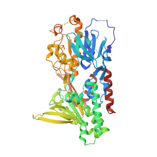
    Yeast Ndi1 is a monotopic alternative NADH dehydrogenase. Its crystal structure in complex with the electron acceptor, ubiquinone, has been determined. However, there has been controversy regarding the ubiquinone binding site. To address these points, we identified the first competitive inhibitor of Ndi1, stigmatellin, along with new mixed-type inhibitors, AC0-12 and myxothiazol, and thereby determined the crystal structures of Ndi1 in complexes with the inhibitors. Two separate binding sites of stigmatellin, STG-1 and STG-2, were observed. The electron density at STG-1, located at the vicinity of the FAD cofactor, further demonstrated two binding modes: STG-1a and STG-1b. AC0-12 and myxothiazol are also located at the vicinity of FAD. The comparison of the binding modes among stigmatellin at STG-1, AC0-12, and myxothiazol revealed a unique position for the aliphatic tail of stigmatellin at STG-1a. Mutations of amino acid residues that interact with this aliphatic tail at STG-1a reduced the affinity of Ndi1 for ubiquinone. In conclusion, the position of the aliphatic tail of stigmatellin at STG-1a provides a structural basis for its competitive inhibition of Ndi1. The inherent binding site of ubiquinone is suggested to overlap with STG-1a that is distinct from the binding site for NADH.


  • Organizational Affiliation

    Department of Cardiovascular Physiology, Faculty of Medicine, Kagawa University, Kita-gun, Kagawa, 761-0793, Japan. tyama@med.kagawa-u.ac.jp.


Macromolecules
Find similar proteins by:  (by identity cutoff)  |  3D Structure
Entity ID: 1
MoleculeChains Sequence LengthOrganismDetailsImage
Rotenone-insensitive NADH-ubiquinone oxidoreductase, mitochondrial484Saccharomyces cerevisiae S288CMutation(s): 0 
Gene Names: NDI1YML120CYM7056.06C
EC: 1.6.5.9
UniProt
Find proteins for P32340 (Saccharomyces cerevisiae (strain ATCC 204508 / S288c))
Explore P32340 
Go to UniProtKB:  P32340
Entity Groups  
Sequence Clusters30% Identity50% Identity70% Identity90% Identity95% Identity100% Identity
UniProt GroupP32340
Sequence Annotations
Expand
  • Reference Sequence
Small Molecules
Ligands 10 Unique
IDChains Name / Formula / InChI Key2D Diagram3D Interactions
FAD
Query on FAD

Download Ideal Coordinates CCD File 
B [auth A]FLAVIN-ADENINE DINUCLEOTIDE
C27 H33 N9 O15 P2
VWWQXMAJTJZDQX-UYBVJOGSSA-N
SMA
Query on SMA

Download Ideal Coordinates CCD File 
AA [auth A],
Z [auth A]
STIGMATELLIN A
C30 H42 O7
UZHDGDDPOPDJGM-WPPYOTIYSA-N
PE4
Query on PE4

Download Ideal Coordinates CCD File 
G [auth A]2-{2-[2-(2-{2-[2-(2-ETHOXY-ETHOXY)-ETHOXY]-ETHOXY}-ETHOXY)-ETHOXY]-ETHOXY}-ETHANOL
C16 H34 O8
PJWQOENWHPEPKI-UHFFFAOYSA-N
P6G
Query on P6G

Download Ideal Coordinates CCD File 
H [auth A]HEXAETHYLENE GLYCOL
C12 H26 O7
IIRDTKBZINWQAW-UHFFFAOYSA-N
MES
Query on MES

Download Ideal Coordinates CCD File 
X [auth A],
Y [auth A]
2-(N-MORPHOLINO)-ETHANESULFONIC ACID
C6 H13 N O4 S
SXGZJKUKBWWHRA-UHFFFAOYSA-N
PG4
Query on PG4

Download Ideal Coordinates CCD File 
M [auth A],
N [auth A]
TETRAETHYLENE GLYCOL
C8 H18 O5
UWHCKJMYHZGTIT-UHFFFAOYSA-N
PGE
Query on PGE

Download Ideal Coordinates CCD File 
Q [auth A],
R [auth A],
T [auth A]
TRIETHYLENE GLYCOL
C6 H14 O4
ZIBGPFATKBEMQZ-UHFFFAOYSA-N
PEG
Query on PEG

Download Ideal Coordinates CCD File 
O [auth A],
P [auth A],
S [auth A]
DI(HYDROXYETHYL)ETHER
C4 H10 O3
MTHSVFCYNBDYFN-UHFFFAOYSA-N
GOL
Query on GOL

Download Ideal Coordinates CCD File 
I [auth A]
J [auth A]
K [auth A]
L [auth A]
U [auth A]
I [auth A],
J [auth A],
K [auth A],
L [auth A],
U [auth A],
V [auth A],
W [auth A]
GLYCEROL
C3 H8 O3
PEDCQBHIVMGVHV-UHFFFAOYSA-N
MG
Query on MG

Download Ideal Coordinates CCD File 
C [auth A],
D [auth A],
E [auth A],
F [auth A]
MAGNESIUM ION
Mg
JLVVSXFLKOJNIY-UHFFFAOYSA-N
Binding Affinity Annotations 
IDSourceBinding Affinity
SMA Binding MOAD:  5YJW IC50: 107 (nM) from 1 assay(s)
Experimental Data & Validation

Experimental Data

  • Method: X-RAY DIFFRACTION
  • Resolution: 1.85 Å
  • R-Value Free: 0.204 
  • R-Value Work: 0.182 
  • R-Value Observed: 0.183 
  • Space Group: C 2 2 21
Unit Cell:
Length ( Å )Angle ( ˚ )
a = 116.67α = 90
b = 128.456β = 90
c = 86.711γ = 90
Software Package:
Software NamePurpose
REFMACrefinement
HKL-2000data reduction
HKL-2000data scaling
MOLREPphasing

Structure Validation

View Full Validation Report


Ligand Structure Quality Assessment 


Entry History & Funding Information

Deposition Data


Funding OrganizationLocationGrant Number
JSPS KAKENHIJapan15K07005

Revision History  (Full details and data files)

  • Version 1.0: 2018-02-14
    Type: Initial release
  • Version 1.1: 2018-02-21
    Changes: Database references
  • Version 1.2: 2023-11-22
    Changes: Data collection, Database references, Derived calculations, Refinement description