5YJM

Human chymase in complex with 7-oxo-3-(phenoxyimino)-1,4-diazepane derivative


Experimental Data Snapshot

  • Method: X-RAY DIFFRACTION
  • Resolution: 1.90 Å
  • R-Value Free: 0.186 
  • R-Value Work: 0.147 
  • R-Value Observed: 0.149 

wwPDB Validation   3D Report Full Report


Ligand Structure Quality Assessment 


This is version 1.2 of the entry. See complete history


Literature

Structure-based design, synthesis, and binding mode analysis of novel and potent chymase inhibitors

Futamura-Takahashi, J.Tanaka, T.Sugawara, H.Iwashita, S.Imajo, S.Oyama, Y.Muto, T.

(2018) Bioorg Med Chem Lett 28: 188-192

  • DOI: https://doi.org/10.1016/j.bmcl.2017.11.031
  • Primary Citation of Related Structures:  
    5YJM, 5YJP

  • PubMed Abstract: 

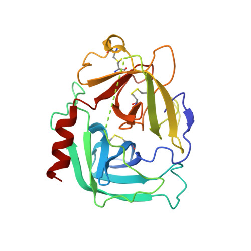
    Based on insight from the X-ray crystal structure of human chymase in complex with compound 1, a lactam carbonyl of the diazepane core was exchanged with O-substituted oxyimino group, leading to amidoxime derivatives. This modification resulted in highly potent chymase inhibitors, such as O-phenylamidoxime 5f. X-ray crystal structure analysis indicated that compound 5f induced movement of the Leu99 and Tyr94 side chains at the S2 site, and the increase in inhibitory activity of O-phenyl amidoxime derivatives suggested that the O-phenyl moiety interacted with the Tyr94 residue. Surface plasmon resonance experiments showed that compound 5f had slower association and dissociation kinetics and the calculated residence time of compound 5f to human chymase was extended compared to that of amide compound 1.


  • Organizational Affiliation

    Asubio Pharma Co., Ltd, 6-4-3 Minatojima-Minamimachi, Chuo-ku, Kobe 650-0047, Japan. Electronic address: futamura.junko.b6@asubio.co.jp.


Macromolecules
Find similar proteins by:  (by identity cutoff)  |  3D Structure
Entity ID: 1
MoleculeChains Sequence LengthOrganismDetailsImage
human chymase226Homo sapiensMutation(s): 0 
UniProt & NIH Common Fund Data Resources
Find proteins for P23946 (Homo sapiens)
Explore P23946 
Go to UniProtKB:  P23946
PHAROS:  P23946
GTEx:  ENSG00000092009 
Entity Groups  
Sequence Clusters30% Identity50% Identity70% Identity90% Identity95% Identity100% Identity
UniProt GroupP23946
Sequence Annotations
Expand
  • Reference Sequence
Small Molecules
Ligands 3 Unique
IDChains Name / Formula / InChI Key2D Diagram3D Interactions
8W3 (Subject of Investigation/LOI)
Query on 8W3

Download Ideal Coordinates CCD File 
E [auth A]2-amino-4-((R)-1-((R,Z)-6-(5-chloro-2-methoxybenzyl)-7-oxo-3-(phenoxyimino)-1,4-diazepane-1-carboxamido)propyl)benzoic acid
C30 H32 Cl N5 O6
LHASZEBEQGPCFM-CJFMBICVSA-N
NAG
Query on NAG

Download Ideal Coordinates CCD File 
B [auth A],
C [auth A]
2-acetamido-2-deoxy-beta-D-glucopyranose
C8 H15 N O6
OVRNDRQMDRJTHS-FMDGEEDCSA-N
ZN
Query on ZN

Download Ideal Coordinates CCD File 
D [auth A]ZINC ION
Zn
PTFCDOFLOPIGGS-UHFFFAOYSA-N
Binding Affinity Annotations 
IDSourceBinding Affinity
8W3 BindingDB:  5YJM Kd: 2.1 (nM) from 1 assay(s)
IC50: 8.9 (nM) from 1 assay(s)
Binding MOAD:  5YJM Kd: 64 (nM) from 1 assay(s)
Experimental Data & Validation

Experimental Data

  • Method: X-RAY DIFFRACTION
  • Resolution: 1.90 Å
  • R-Value Free: 0.186 
  • R-Value Work: 0.147 
  • R-Value Observed: 0.149 
  • Space Group: P 43
Unit Cell:
Length ( Å )Angle ( ˚ )
a = 75.089α = 90
b = 75.089β = 90
c = 49.75γ = 90
Software Package:
Software NamePurpose
REFMACrefinement
HKL-2000data reduction
HKL-2000data scaling
AMoREphasing

Structure Validation

View Full Validation Report


Ligand Structure Quality Assessment 


Entry History 

Deposition Data

  • Released Date: 2017-12-27 
  • Deposition Author(s): Sugawara, H.

Revision History  (Full details and data files)

  • Version 1.0: 2017-12-27
    Type: Initial release
  • Version 1.1: 2018-01-17
    Changes: Database references
  • Version 1.2: 2020-07-29
    Type: Remediation
    Reason: Carbohydrate remediation
    Changes: Data collection, Derived calculations, Structure summary