5YGY | pdb_00005ygy

Crystal Structure of BACE1 in complex with (S)-N-(3-(2-amino-6-(fluoromethyl)-4 -methyl-4H-1,3-oxazin-4-yl)-4-fluorophenyl)-5-cyanopicolinamide


Experimental Data Snapshot

  • Method: X-RAY DIFFRACTION
  • Resolution: 2.30 Å
  • R-Value Free: 
    0.235 (Depositor), 0.239 (DCC) 
  • R-Value Work: 
    0.191 (Depositor), 0.195 (DCC) 
  • R-Value Observed: 
    0.196 (Depositor) 

Starting Model: experimental
View more details

wwPDB Validation 3D Report Full Report

Validation slider image for 5YGY

Ligand Structure Quality Assessment 


This is version 1.4 of the entry. See complete history

Literature

Rational Design of Novel 1,3-Oxazine Based beta-Secretase (BACE1) Inhibitors: Incorporation of a Double Bond To Reduce P-gp Efflux Leading to Robust A beta Reduction in the Brain

Fuchino, K.Mitsuoka, Y.Masui, M.Kurose, N.Yoshida, S.Komano, K.Yamamoto, T.Ogawa, M.Unemura, C.Hosono, M.Ito, H.Sakaguchi, G.Ando, S.Ohnishi, S.Kido, Y.Fukushima, T.Miyajima, H.Hiroyama, S.Koyabu, K.Dhuyvetter, D.Borghys, H.Gijsen, H.J.M.Yamano, Y.Iso, Y.Kusakabe, K.I.

(2018) J Med Chem 61: 5122-5137

  • DOI: https://doi.org/10.1021/acs.jmedchem.8b00002
  • Primary Citation Related Structures: 
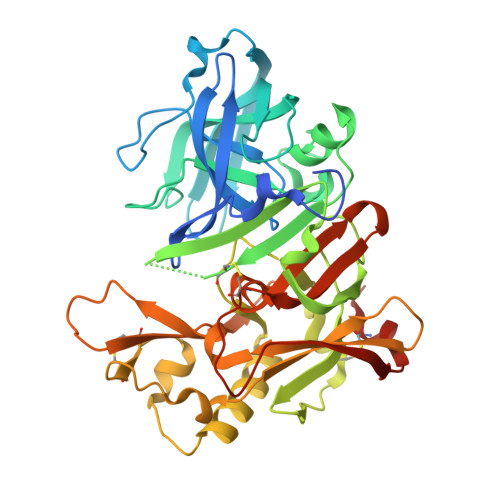
    5YGY

  • PubMed Abstract: 

    Accumulation of Aβ peptides is a hallmark of Alzheimer's disease (AD) and is considered a causal factor in the pathogenesis of AD. β-Secretase (BACE1) is a key enzyme responsible for producing Aβ peptides, and thus agents that inhibit BACE1 should be beneficial for disease-modifying treatment of AD. Here we describe the discovery and optimization of novel oxazine-based BACE1 inhibitors by lowering amidine basicity with the incorporation of a double bond to improve brain penetration. Starting from a 1,3-dihydrooxazine lead 6 identified by a hit-to-lead SAR following HTS, we adopted a p K a lowering strategy to reduce the P-gp efflux and the high hERG potential leading to the discovery of 15 that produced significant Aβ reduction with long duration in pharmacodynamic models and exhibited wide safety margins in cardiovascular safety models. This compound improved the brain-to-plasma ratio relative to 6 by reducing P-gp recognition, which was demonstrated by a P-gp knockout mouse model.

Macromolecule Content 

  • Total Structure Weight: 47.69 kDa 
  • Atom Count: 3,190 
  • Modeled Residue Count: 381 
  • Deposited Residue Count: 416 
  • Unique protein chains: 1

Macromolecules

Find similar proteins by:|  3D Structure
Entity ID: 1
MoleculeChains  Sequence LengthOrganismDetailsImage
Beta-secretase 1416Homo sapiensMutation(s): 0 
Gene Names: BACE1BACEKIAA1149
EC: 3.4.23.46
UniProt & NIH Common Fund Data Resources
Find proteins for P56817 (Homo sapiens)
Explore P56817 
Go to UniProtKB:  P56817
PHAROS:  P56817
GTEx:  ENSG00000186318 
Entity Groups
Sequence Clusters30% Identity50% Identity70% Identity90% Identity95% Identity100% Identity
UniProt GroupP56817
Sequence Annotations
Expand
Reference Sequence

Small Molecules

Ligands 3 Unique
IDChains Name / Formula / InChI Key2D Diagram3D Interactions
0B6
(Subject of Investigation/LOI)

Query on 0B6


Download:Ideal Coordinates CCD File
J [auth A]~{N}-[3-[(4~{S})-2-azanyl-6-(fluoranylmethyl)-4-methyl-1,3-oxazin-4-yl]-4-fluoranyl-phenyl]-5-cyano-pyridine-2-carboxamide
C19 H15 F2 N5 O2
ORHZQSYKGHHDOP-IBGZPJMESA-N
IOD

Query on IOD


Download:Ideal Coordinates CCD File
B [auth A],
C [auth A],
D [auth A],
E [auth A],
F [auth A]
IODIDE ION
I
XMBWDFGMSWQBCA-UHFFFAOYSA-M
GOL

Query on GOL


Download:Ideal Coordinates CCD File
G [auth A],
H [auth A],
I [auth A]
GLYCEROL
C3 H8 O3
PEDCQBHIVMGVHV-UHFFFAOYSA-N
Binding Affinity Annotations 
IDSourceBinding Affinity
0B6 BindingDB:  5YGY IC50: min: 3.6, max: 59 (nM) from 2 assay(s)

Experimental Data & Validation

Experimental Data

  • Method: X-RAY DIFFRACTION
  • Resolution: 2.30 Å
  • R-Value Free:  0.235 (Depositor), 0.239 (DCC) 
  • R-Value Work:  0.191 (Depositor), 0.195 (DCC) 
  • R-Value Observed: 0.196 (Depositor) 
Space Group: P 61 2 2
Unit Cell:
Length ( Å )Angle ( ˚ )
a = 102.149α = 90
b = 102.149β = 90
c = 171.125γ = 120
Software Package:
Software NamePurpose
HKL-2000data processing
HKL-2000data scaling
MOLREPphasing
REFMACrefinement
PDB_EXTRACTdata extraction

Structure Validation

View Full Validation Report


Ligand Structure Quality Assessment 


Entry History 

Revision History  (Full details and data files)

  • Version 1.0: 2018-05-23
    Type: Initial release
  • Version 1.1: 2018-05-30
    Changes: Data collection, Database references
  • Version 1.2: 2018-07-11
    Changes: Data collection, Database references
  • Version 1.3: 2023-11-22
    Changes: Data collection, Database references, Refinement description
  • Version 1.4: 2024-11-13
    Changes: Structure summary