5YFZ

Plasmodium vivax SHMT bound with PLP-glycine and S-GS626


Experimental Data Snapshot

  • Method: X-RAY DIFFRACTION
  • Resolution: 2.16 Å
  • R-Value Free: 0.333 
  • R-Value Work: 0.263 
  • R-Value Observed: 0.270 

wwPDB Validation   3D Report Full Report


Ligand Structure Quality Assessment 


This is version 1.1 of the entry. See complete history


Literature

Potent Inhibitors of Plasmodial Serine Hydroxymethyltransferase (SHMT) Featuring a Spirocyclic Scaffold

Schwertz, G.Witschel, M.C.Rottmann, M.Leartsakulpanich, U.Chitnumsub, P.Jaruwat, A.Amornwatcharapong, W.Ittarat, W.Schafer, A.Aponte, R.A.Trapp, N.Chaiyen, P.Diederich, F.

(2018) ChemMedChem 13: 931-943

  • DOI: https://doi.org/10.1002/cmdc.201800053
  • Primary Citation of Related Structures:  
    5YFZ, 5YG2, 5YG3, 5YG4

  • PubMed Abstract: 

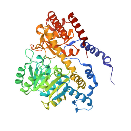
    With the discovery that serine hydroxymethyltransferase (SHMT) is a druggable target for antimalarials, the aim of this study was to design novel inhibitors of this key enzyme in the folate biosynthesis cycle. Herein, 19 novel spirocyclic ligands based on either 2-indolinone or dihydroindene scaffolds and featuring a pyrazolopyran core are reported. Strong target affinities for Plasmodium falciparum (Pf) SHMT (14-76 nm) and cellular potencies in the low nanomolar range (165-334 nm) were measured together with interesting selectivity against human cytosolic SHMT1 (hSHMT1). Four co-crystal structures with Plasmodium vivax (Pv) SHMT solved at 2.2-2.4 Å resolution revealed the key role of the vinylogous cyanamide for anchoring ligands within the active site. The spirocyclic motif in the molecules enforces the pyrazolopyran core to adopt a substantially more curved conformation than that of previous non-spirocyclic analogues. Finally, solvation of the spirocyclic lactam ring of the receptor-bound ligands is discussed.


  • Organizational Affiliation

    Laboratorium für Organische Chemie, ETH Zürich, Vladimir-Prelog-Weg 3, 8093, Zürich, Switzerland.


Macromolecules
Find similar proteins by:  (by identity cutoff)  |  3D Structure
Entity ID: 1
MoleculeChains Sequence LengthOrganismDetailsImage
Serine hydroxymethyltransferase
A, B, C
442Plasmodium vivaxMutation(s): 0 
Gene Names: PVC01_140059500PVP01_1453700PVT01_140059000
EC: 2.1.2.1
UniProt
Find proteins for A0A1G4H5I1 (Plasmodium vivax)
Explore A0A1G4H5I1 
Go to UniProtKB:  A0A1G4H5I1
Entity Groups  
Sequence Clusters30% Identity50% Identity70% Identity90% Identity95% Identity100% Identity
UniProt GroupA0A1G4H5I1
Sequence Annotations
Expand
  • Reference Sequence
Small Molecules
Ligands 3 Unique
IDChains Name / Formula / InChI Key2D Diagram3D Interactions
8UO
Query on 8UO

Download Ideal Coordinates CCD File 
E [auth A],
H [auth B],
J [auth C]
2-[1-[3-[(4~{S})-6-azanyl-5-cyano-3-methyl-4-propan-2-yl-2~{H}-pyrano[2,3-c]pyrazol-4-yl]-5-fluoranyl-phenyl]piperidin-4-yl]ethanoic acid
C24 H28 F N5 O3
LNTOYLXMWSSXMA-DEOSSOPVSA-N
PLG
Query on PLG

Download Ideal Coordinates CCD File 
D [auth A],
G [auth B],
I [auth C]
N-GLYCINE-[3-HYDROXY-2-METHYL-5-PHOSPHONOOXYMETHYL-PYRIDIN-4-YL-METHANE]
C10 H15 N2 O7 P
FEVQWBMNLWUBTF-UHFFFAOYSA-N
CL
Query on CL

Download Ideal Coordinates CCD File 
F [auth A],
K [auth C]
CHLORIDE ION
Cl
VEXZGXHMUGYJMC-UHFFFAOYSA-M
Experimental Data & Validation

Experimental Data

  • Method: X-RAY DIFFRACTION
  • Resolution: 2.16 Å
  • R-Value Free: 0.333 
  • R-Value Work: 0.263 
  • R-Value Observed: 0.270 
  • Space Group: C 1 2 1
Unit Cell:
Length ( Å )Angle ( ˚ )
a = 101.875α = 90
b = 58.796β = 89.99
c = 235.728γ = 90
Software Package:
Software NamePurpose
REFMACrefinement
SCALEPACKdata scaling
PDB_EXTRACTdata extraction
PHASERphasing

Structure Validation

View Full Validation Report


Ligand Structure Quality Assessment 


Entry History 

Deposition Data

Revision History  (Full details and data files)

  • Version 1.0: 2018-06-06
    Type: Initial release
  • Version 1.1: 2023-11-22
    Changes: Data collection, Database references, Refinement description