Structure, interactions and action ofMycobacterium tuberculosis3-hydroxyisobutyric acid dehydrogenase.
Srikalaivani, R., Singh, A., Vijayan, M., Surolia, A.(2018) Biochem J 475: 2457-2471
- PubMed: 29959185 Search on PubMed
- DOI: https://doi.org/10.1042/BCJ20180271
- Primary Citation Related Structures:
5Y8G, 5Y8H, 5Y8I, 5Y8J, 5Y8K, 5Y8L, 5Y8M, 5Y8N, 5Y8O, 5Y8P - PubMed Abstract:
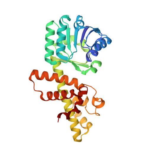
Biochemical and crystallographic studies on Mycobacterium tuberculosis 3-hydroxyisobutyric acid dehydrogenase ( Mt HIBADH), a member of the 3-hydroxyacid dehydrogenase superfamily, have been carried out. Gel filtration and blue native PAGE of Mt HIBADH show that the enzyme is a dimer. The enzyme preferentially uses NAD + as the cofactor and is specific to S -hydroxyisobutyric acid (HIBA). It can also use R -HIBA, l-serine and 3-hydroxypropanoic acid (3-HP) as substrates, but with much less efficiency. The pH optimum for activity is ∼11. Structures of the native enzyme, the holoenzyme, binary complexes with NAD + , S -HIBA, R -HIBA, l-serine and 3-HP and ternary complexes involving the substrates and NAD + have been determined. None of the already known structures of HIBADH contain a substrate molecule at the binding site. The structures reported here provide for the first time, among other things, a clear indication of the location and interactions of the substrates at the active site. They also define the entrance of the substrates to the active site region. The structures provide information on the role of specific residues at the active site and the entrance. The results obtained from crystal structures are consistent with solution studies including mutational analysis. They lead to the proposal of a plausible mechanism of the action of the enzyme.
- Molecular Biophysics Unit, Indian Institute of Science, Bangalore, India.
Organizational Affiliation: