5Y7Y

Crystal structure of AhRR/ARNT complex


Experimental Data Snapshot

  • Method: X-RAY DIFFRACTION
  • Resolution: 2.40 Å
  • R-Value Free: 0.268 
  • R-Value Work: 0.234 
  • R-Value Observed: 0.235 

wwPDB Validation   3D Report Full Report


This is version 1.1 of the entry. See complete history


Literature

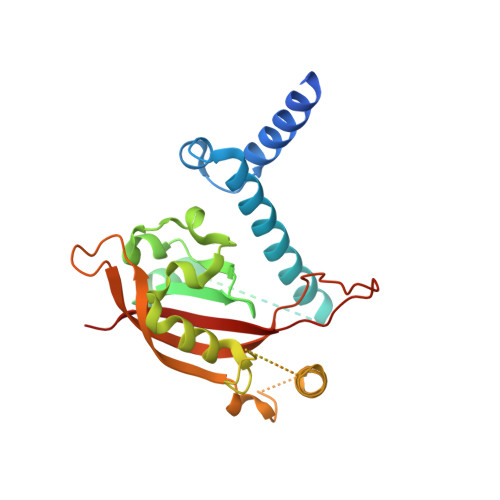
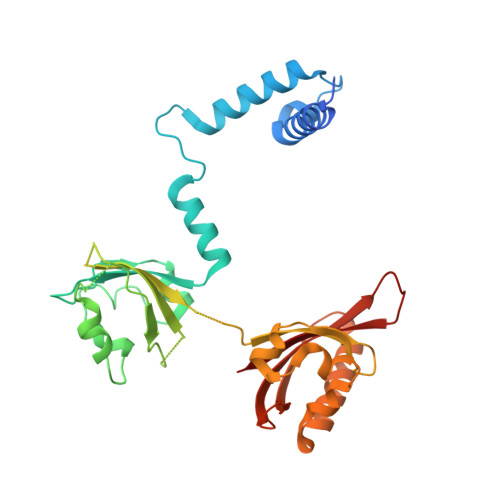
The crystal structure of the AhRR-ARNT heterodimer reveals the structural basis of the repression of AhR-mediated transcription.

Sakurai, S.Shimizu, T.Ohto, U.

(2017) J Biol Chem 292: 17609-17616

  • DOI: https://doi.org/10.1074/jbc.M117.812974
  • Primary Citation of Related Structures:  
    5Y7Y

  • PubMed Abstract: 
  • 2,3,7,8-Tetrachlorodibenzo- p -dioxin and related compounds are extraordinarily potent environmental toxic pollutants. Most of the 2,3,7,8-tetrachlorodibenzo- p -dioxin toxicities are mediated by aryl hydrocarbon receptor (AhR), a ligand-dependent transcription factor belonging to the basic helix-loop-helix (bHLH) Per-ARNT-Sim (PAS) family ...

    2,3,7,8-Tetrachlorodibenzo- p -dioxin and related compounds are extraordinarily potent environmental toxic pollutants. Most of the 2,3,7,8-tetrachlorodibenzo- p -dioxin toxicities are mediated by aryl hydrocarbon receptor (AhR), a ligand-dependent transcription factor belonging to the basic helix-loop-helix (bHLH) Per-ARNT-Sim (PAS) family. Upon ligand binding, AhR forms a heterodimer with AhR nuclear translocator (ARNT) and induces the expression of genes involved in various biological responses. One of the genes induced by AhR encodes AhR repressor (AhRR), which also forms a heterodimer with ARNT and represses the activation of AhR-dependent transcription. The control of AhR activation is critical for managing AhR-mediated diseases, but the mechanisms by which AhRR represses AhR activation remain poorly understood, because of the lack of structural information. Here, we determined the structure of the AhRR-ARNT heterodimer by X-ray crystallography, which revealed an asymmetric intertwined domain organization presenting structural features that are both conserved and distinct among bHLH-PAS family members. The structures of AhRR-ARNT and AhR-ARNT were similar in the bHLH-PAS-A region, whereas the PAS-B of ARNT in the AhRR-ARNT complex exhibited a different domain arrangement in this family reported so far. The structure clearly disclosed that AhRR competitively represses AhR binding to ARNT and target DNA and further suggested the existence of an AhRR-ARNT-specific repression mechanism. This study provides a structural basis for understanding the mechanism by which AhRR represses AhR-mediated gene transcription.


    Organizational Affiliation

    From the Graduate School of Pharmaceutical Sciences, University of Tokyo, Tokyo 113-0033, Japan and umeji@mol.f.u-tokyo.ac.jp.


Macromolecules
Find similar proteins by:  (by identity cutoff)  |  3D Structure
Entity ID: 1
MoleculeChainsSequence LengthOrganismDetailsImage
Aryl hydrocarbon receptor repressorA258Homo sapiensMutation(s): 0 
Gene Names: AHRRBHLHE77KIAA1234
UniProt & NIH Common Fund Data Resources
Find proteins for A9YTQ3 (Homo sapiens)
Explore A9YTQ3 
Go to UniProtKB:  A9YTQ3
PHAROS:  A9YTQ3
Entity Groups  
Sequence Clusters30% Identity50% Identity70% Identity90% Identity95% Identity100% Identity
UniProt GroupA9YTQ3
Protein Feature View
Expand
  • Reference Sequence
Find similar proteins by:  (by identity cutoff)  |  3D Structure
Entity ID: 2
MoleculeChainsSequence LengthOrganismDetailsImage
Aryl hydrocarbon receptor nuclear translocatorB311Bos taurusMutation(s): 0 
Gene Names: ARNT
UniProt
Find proteins for Q9BE97 (Bos taurus)
Explore Q9BE97 
Go to UniProtKB:  Q9BE97
Entity Groups  
Sequence Clusters30% Identity50% Identity70% Identity90% Identity95% Identity100% Identity
UniProt GroupQ9BE97
Protein Feature View
Expand
  • Reference Sequence
Small Molecules
Ligands 1 Unique
IDChainsName / Formula / InChI Key2D Diagram3D Interactions
GOL
Query on GOL

Download Ideal Coordinates CCD File 
C [auth A],
D [auth A],
E [auth B]
GLYCEROL
C3 H8 O3
PEDCQBHIVMGVHV-UHFFFAOYSA-N
 Ligand Interaction
Experimental Data & Validation

Experimental Data

  • Method: X-RAY DIFFRACTION
  • Resolution: 2.40 Å
  • R-Value Free: 0.268 
  • R-Value Work: 0.234 
  • R-Value Observed: 0.235 
  • Space Group: P 41
Unit Cell:
Length ( Å )Angle ( ˚ )
a = 78.362α = 90
b = 78.362β = 90
c = 129.784γ = 90
Software Package:
Software NamePurpose
REFMACrefinement
HKL-2000data reduction
HKL-2000data scaling
PHASERphasing

Structure Validation

View Full Validation Report


Entry History 

Deposition Data

Revision History  (Full details and data files)

  • Version 1.0: 2017-09-20
    Type: Initial release
  • Version 1.1: 2018-02-21
    Changes: Database references