5Y6A

Crystal structure of the anti-CRISPR protein, AcrIIA1


Experimental Data Snapshot

  • Method: X-RAY DIFFRACTION
  • Resolution: 2.00 Å
  • R-Value Free: 0.228 
  • R-Value Work: 0.183 
  • R-Value Observed: 0.185 

wwPDB Validation   3D Report Full Report


This is version 1.3 of the entry. See complete history


Literature

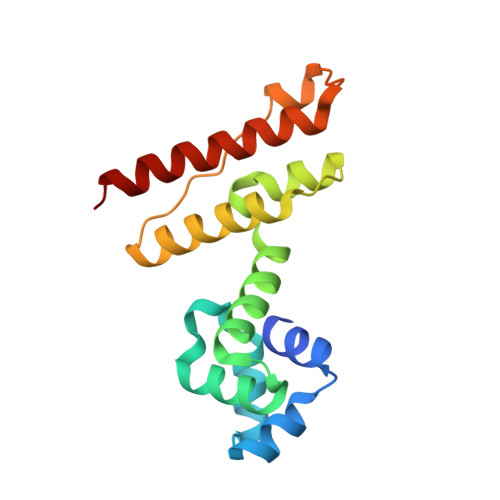
Crystal structure of an anti-CRISPR protein, AcrIIA1

Ka, D.An, S.Y.Suh, J.Y.Bae, E.

(2018) Nucleic Acids Res 46: 485-492

  • DOI: https://doi.org/10.1093/nar/gkx1181
  • Primary Citation of Related Structures:  
    5Y69, 5Y6A

  • PubMed Abstract: 

    Clustered regularly interspaced short palindromic repeats (CRISPRs) and CRISPR-associated (Cas) proteins provide bacteria with RNA-based adaptive immunity against phage infection. To counteract this defense mechanism, phages evolved anti-CRISPR (Acr) proteins that inactivate the CRISPR-Cas systems. AcrIIA1, encoded by Listeria monocytogenes prophages, is the most prevalent among the Acr proteins targeting type II-A CRISPR-Cas systems and has been used as a marker to identify other Acr proteins. Here, we report the crystal structure of AcrIIA1 and its RNA-binding affinity. AcrIIA1 forms a dimer with a novel two helical-domain architecture. The N-terminal domain of AcrIIA1 exhibits a helix-turn-helix motif similar to transcriptional factors. When overexpressed in Escherichia coli, AcrIIA1 associates with RNAs, suggesting that AcrIIA1 functions via nucleic acid recognition. Taken together, the unique structural and functional features of AcrIIA1 suggest its distinct mode of Acr activity, expanding the diversity of the inhibitory mechanisms employed by Acr proteins.


  • Organizational Affiliation

    Department of Agricultural Biotechnology, Seoul National University, Seoul 08826, Korea.


Macromolecules
Find similar proteins by:  (by identity cutoff)  |  3D Structure
Entity ID: 1
MoleculeChains Sequence LengthOrganismDetailsImage
chain A and B
A, B
151Listeria monocytogenes J0161Mutation(s): 0 
Gene Names: LMOG_03146
UniProt
Find proteins for A8ATW7 (Listeria phage A006)
Explore A8ATW7 
Go to UniProtKB:  A8ATW7
Entity Groups  
Sequence Clusters30% Identity50% Identity70% Identity90% Identity95% Identity100% Identity
UniProt GroupA8ATW7
Sequence Annotations
Expand
  • Reference Sequence
Experimental Data & Validation

Experimental Data

  • Method: X-RAY DIFFRACTION
  • Resolution: 2.00 Å
  • R-Value Free: 0.228 
  • R-Value Work: 0.183 
  • R-Value Observed: 0.185 
  • Space Group: C 1 2 1
Unit Cell:
Length ( Å )Angle ( ˚ )
a = 127.435α = 90
b = 55.42β = 96.01
c = 46.504γ = 90
Software Package:
Software NamePurpose
PHENIXrefinement
HKL-2000data processing
HKL-2000data scaling
PHENIXphasing
HKL-2000data reduction

Structure Validation

View Full Validation Report


Entry History & Funding Information

Deposition Data


Funding OrganizationLocationGrant Number
Rural Development AdministrationKorea, Republic OfPJ01111201
the Basic Science Research Program through the National Research Foundation of Korea funded by the Ministry of Education, Science and TechnologyKorea, Republic OfNRF-2016R1D1A1A09916821

Revision History  (Full details and data files)

  • Version 1.0: 2017-11-29
    Type: Initial release
  • Version 1.1: 2017-12-13
    Changes: Database references
  • Version 1.2: 2018-01-24
    Changes: Database references
  • Version 1.3: 2023-11-22
    Changes: Data collection, Database references, Refinement description