5Y17

CATPO mutant - E316F


Experimental Data Snapshot

  • Method: X-RAY DIFFRACTION
  • Resolution: 2.30 Å
  • R-Value Free: 0.220 
  • R-Value Work: 0.178 
  • R-Value Observed: 0.180 

wwPDB Validation   3D Report Full Report


Ligand Structure Quality Assessment 


This is version 1.3 of the entry. See complete history


Literature

Identification of the site of oxidase substrate binding in Scytalidium thermophilum catalase.

Yuzugullu Karakus, Y.Goc, G.Balci, S.Yorke, B.A.Trinh, C.H.McPherson, M.J.Pearson, A.R.

(2018) Acta Crystallogr D Struct Biol 74: 979-985

  • DOI: https://doi.org/10.1107/S2059798318010628
  • Primary Citation of Related Structures:  
    5XVZ, 5XY4, 5Y17, 5ZZ1

  • PubMed Abstract: 

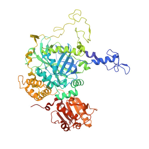
    The catalase from Scytalidium thermophilum is a homotetramer containing a heme d in each active site. Although the enzyme has a classical monofunctional catalase fold, it also possesses oxidase activity towards a number of small organics, including catechol and phenol. In order to further investigate this, the crystal structure of the complex of the catalase with the classical catalase inhibitor 3-amino-1,2,4-triazole (3TR) was determined at 1.95 Å resolution. Surprisingly, no binding to the heme site was observed; instead, 3TR occupies a binding site corresponding to the NADPH-binding pocket in mammalian catalases at the entrance to a lateral channel leading to the heme. Kinetic analysis of site-directed mutants supports the assignment of this pocket as the binding site for oxidase substrates.


  • Organizational Affiliation

    Department of Biology, Kocaeli University, Umuttepe, 41380 Kocaeli, Turkey.


Macromolecules
Find similar proteins by:  (by identity cutoff)  |  3D Structure
Entity ID: 1
MoleculeChains Sequence LengthOrganismDetailsImage
Catalase
A, B, C, D
678Mycothermus thermophilusMutation(s): 1 
EC: 1.11.1.6
UniProt
Find proteins for M4GGR7 (Mycothermus thermophilus)
Explore M4GGR7 
Go to UniProtKB:  M4GGR7
Entity Groups  
Sequence Clusters30% Identity50% Identity70% Identity90% Identity95% Identity100% Identity
UniProt GroupM4GGR7
Sequence Annotations
Expand
  • Reference Sequence
Small Molecules
Ligands 2 Unique
IDChains Name / Formula / InChI Key2D Diagram3D Interactions
HDD
Query on HDD

Download Ideal Coordinates CCD File 
E [auth A],
J [auth B],
M [auth C],
P [auth D]
CIS-HEME D HYDROXYCHLORIN GAMMA-SPIROLACTONE
C34 H32 Fe N4 O5
UMGOPAWIGKFTRK-QQDQPIDJSA-N
CA
Query on CA

Download Ideal Coordinates CCD File 
F [auth A]
G [auth A]
H [auth A]
I [auth A]
K [auth B]
F [auth A],
G [auth A],
H [auth A],
I [auth A],
K [auth B],
L [auth B],
N [auth C],
O [auth C],
Q [auth D],
R [auth D]
CALCIUM ION
Ca
BHPQYMZQTOCNFJ-UHFFFAOYSA-N
Experimental Data & Validation

Experimental Data

  • Method: X-RAY DIFFRACTION
  • Resolution: 2.30 Å
  • R-Value Free: 0.220 
  • R-Value Work: 0.178 
  • R-Value Observed: 0.180 
  • Space Group: I 1 2 1
Unit Cell:
Length ( Å )Angle ( ˚ )
a = 125.733α = 90
b = 120.87β = 101.97
c = 183.804γ = 90
Software Package:
Software NamePurpose
REFMACrefinement
PDB_EXTRACTdata extraction
XDSdata reduction
Aimlessdata scaling
MOLREPphasing

Structure Validation

View Full Validation Report


Ligand Structure Quality Assessment 


Entry History & Funding Information

Deposition Data


Funding OrganizationLocationGrant Number
TUBITAKTurkey113Z744

Revision History  (Full details and data files)

  • Version 1.0: 2018-07-25
    Type: Initial release
  • Version 1.1: 2018-08-01
    Changes: Data collection, Experimental preparation
  • Version 1.2: 2019-07-31
    Changes: Data collection, Database references
  • Version 1.3: 2023-11-22
    Changes: Data collection, Database references, Derived calculations, Refinement description