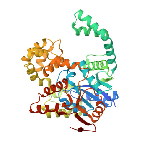
5Y0Q

Crystal structure of BsTmcAL bound with AMPCPP


Experimental Data Snapshot

  • Method: X-RAY DIFFRACTION
  • Resolution: 2.10 Å
  • R-Value Free: 0.243 
  • R-Value Work: 0.202 
  • R-Value Observed: 0.205 

Starting Model: experimental
View more details

wwPDB Validation   3D Report Full Report


Ligand Structure Quality Assessment 


This is version 1.2 of the entry. See complete history


Literature

Acetate-dependent tRNA acetylation required for decoding fidelity in protein synthesis.

Taniguchi, T.Miyauchi, K.Sakaguchi, Y.Yamashita, S.Soma, A.Tomita, K.Suzuki, T.

(2018) Nat Chem Biol 14: 1010-1020

  • DOI: https://doi.org/10.1038/s41589-018-0119-z
  • Primary Citation of Related Structures:  
    5Y0N, 5Y0O, 5Y0P, 5Y0Q, 5Y0R, 5Y0S, 5Y0T

  • PubMed Abstract: 

    Modification of tRNA anticodons plays a critical role in ensuring accurate translation. N 4 -acetylcytidine (ac 4 C) is present at the anticodon first position (position 34) of bacterial elongator tRNA Met . Herein, we identified Bacillus subtilis ylbM (renamed tmcAL) as a novel gene responsible for ac 4 C34 formation. Unlike general acetyltransferases that use acetyl-CoA, TmcAL activates an acetate ion to form acetyladenylate and then catalyzes ac 4 C34 formation through a mechanism similar to tRNA aminoacylation. The crystal structure of TmcAL with an ATP analog reveals the molecular basis of ac 4 C34 formation. The ΔtmcAL strain displayed a cold-sensitive phenotype and a strong genetic interaction with tilS that encodes the enzyme responsible for synthesizing lysidine (L) at position 34 of tRNA Ile to facilitate AUA decoding. Mistranslation of the AUA codon as Met in the ΔtmcAL strain upon tilS repression suggests that ac 4 C34 modification of tRNA Met and L34 modification of tRNA Ile act cooperatively to prevent misdecoding of the AUA codon.


  • Organizational Affiliation

    Department of Chemistry and Biotechnology, Graduate School of Engineering, University of Tokyo, Bunkyo-ku, Tokyo, Japan.


Macromolecules
Find similar proteins by:  (by identity cutoff)  |  3D Structure
Entity ID: 1
MoleculeChains Sequence LengthOrganismDetailsImage
UPF0348 protein B4417_3650
A, B
434Bacillus subtilisMutation(s): 0 
Gene Names: B4417_3650
UniProt
Find proteins for O34513 (Bacillus subtilis (strain 168))
Explore O34513 
Go to UniProtKB:  O34513
Entity Groups  
Sequence Clusters30% Identity50% Identity70% Identity90% Identity95% Identity100% Identity
UniProt GroupO34513
Sequence Annotations
Expand
  • Reference Sequence
Small Molecules
Ligands 1 Unique
IDChains Name / Formula / InChI Key2D Diagram3D Interactions
APC
Query on APC

Download Ideal Coordinates CCD File 
C [auth A],
D [auth B]
DIPHOSPHOMETHYLPHOSPHONIC ACID ADENOSYL ESTER
C11 H18 N5 O12 P3
CAWZRIXWFRFUQB-IOSLPCCCSA-N
Experimental Data & Validation

Experimental Data

  • Method: X-RAY DIFFRACTION
  • Resolution: 2.10 Å
  • R-Value Free: 0.243 
  • R-Value Work: 0.202 
  • R-Value Observed: 0.205 
  • Space Group: P 21 21 21
Unit Cell:
Length ( Å )Angle ( ˚ )
a = 67.995α = 90
b = 68.013β = 90
c = 211.672γ = 90
Software Package:
Software NamePurpose
XSCALEdata scaling
PHENIXrefinement
PDB_EXTRACTdata extraction
XDSdata reduction
PHASERphasing

Structure Validation

View Full Validation Report


Ligand Structure Quality Assessment 


Entry History 

Deposition Data

Revision History  (Full details and data files)

  • Version 1.0: 2018-07-18
    Type: Initial release
  • Version 1.1: 2018-12-26
    Changes: Data collection, Database references, Structure summary
  • Version 1.2: 2023-11-22
    Changes: Data collection, Database references, Refinement description